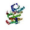
 Movie
Movie Controller


+ Open data

- Basic information
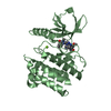
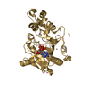
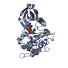
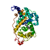
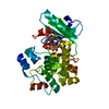
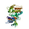
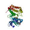
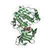
| Entry | Database: PDB / ID: 5yv9 | ||||||
|---|---|---|---|---|---|---|---|
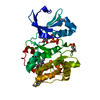
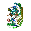
| Title | Structure of CaMKK2 in complex with CKI-009 | ||||||
 Components Components | Calcium/calmodulin-dependent protein kinase kinase 2 | ||||||
 Keywords Keywords | TRANSFERASE / ATP-BINDING / KINASE / SERINE/THREONINE-PROTEIN KINASE / PROTEIN-INHIBITOR COMPLEX | ||||||
| Function / homology |  Function and homology information Function and homology informationregulation of protein kinase activity / positive regulation of autophagy of mitochondrion / Ca2+/calmodulin-dependent protein kinase / CAMKK-AMPK signaling cascade / calcium/calmodulin-dependent protein kinase activity / CREB1 phosphorylation through the activation of CaMKII/CaMKK/CaMKIV cascasde / CaMK IV-mediated phosphorylation of CREB / Activation of RAC1 downstream of NMDARs / Activation of AMPK downstream of NMDARs / cellular response to reactive oxygen species ...regulation of protein kinase activity / positive regulation of autophagy of mitochondrion / Ca2+/calmodulin-dependent protein kinase / CAMKK-AMPK signaling cascade / calcium/calmodulin-dependent protein kinase activity / CREB1 phosphorylation through the activation of CaMKII/CaMKK/CaMKIV cascasde / CaMK IV-mediated phosphorylation of CREB / Activation of RAC1 downstream of NMDARs / Activation of AMPK downstream of NMDARs / cellular response to reactive oxygen species / calcium-mediated signaling / protein autophosphorylation / MAPK cascade / protein tyrosine kinase activity / protein phosphorylation / calmodulin binding / neuron projection / protein serine kinase activity / protein serine/threonine kinase activity / calcium ion binding / positive regulation of DNA-templated transcription / nucleoplasm / ATP binding / cytosol Similarity search - Function | ||||||
| Biological species |  Homo sapiens (human) Homo sapiens (human) | ||||||
| Method |  X-RAY DIFFRACTION / SYNCHROTRON / MOLECULAR REPLACEMENT / Resolution: 2.53 Å X-RAY DIFFRACTION / SYNCHROTRON / MOLECULAR REPLACEMENT / Resolution: 2.53 Å | ||||||
 Authors Authors | Niwa, H. / Handa, N. / Yokoyama, S. | ||||||
 Citation Citation | Journal: J.Mol.Graph.Model. / Year: 2020 Title: Protein ligand interaction analysis against new CaMKK2 inhibitors by use of X-ray crystallography and the fragment molecular orbital (FMO) method. Authors: Takaya, D. / Niwa, H. / Mikuni, J. / Nakamura, K. / Handa, N. / Tanaka, A. / Yokoyama, S. / Honma, T. | ||||||
| History |
|
- Structure visualization
| Structure viewer | Molecule: MolmilJmol/JSmol |
|---|
- Downloads & links
-Download
| PDBx/mmCIF format | 5yv9.cif.gz | 126.2 KB | Display | PDBx/mmCIF format |
|---|---|---|---|---|
| PDB format | pdb5yv9.ent.gz | 96.1 KB | Display | PDB format |
| PDBx/mmJSON format | 5yv9.json.gz | Tree view | PDBx/mmJSON format | |
| Others |  Other downloads Other downloads |
-Validation report
| Arichive directory | https://data.pdbj.org/pub/pdb/validation_reports/yv/5yv9ftp://data.pdbj.org/pub/pdb/validation_reports/yv/5yv9 | HTTPS FTP |
|---|
-Related structure data
| Related structure data |  5yv8C  5yvaC  5yvbC  5yvcC  2zv2S S: Starting model for refinement C: citing same article ( |
|---|---|
| Similar structure data |


-Links
 PDBj
PDBj


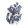
- Assembly
| Deposited unit | 
| ||||||||
|---|---|---|---|---|---|---|---|---|---|
| 1 |
| ||||||||
| Unit cell |
|
-Components
| #1: Protein | Mass: 33711.637 Da / Num. of mol.: 1 Source method: isolated from a genetically manipulated source Source: (gene. exp.) Homo sapiens (human) / Gene: CAMKK2, CAMKKB, KIAA0787 / Plasmid: PX070406-05 / Production host: CELL-FREE SYNTHESIS (others)References: UniProt: Q96RR4, Ca2+/calmodulin-dependent protein kinase | ||||||
|---|---|---|---|---|---|---|---|
| #2: Chemical | ChemComp-91O /   Mass: 361.776 Da / Num. of mol.: 1 / Source method: obtained synthetically / Formula: C18H16ClNO5 / Feature type: SUBJECT OF INVESTIGATION Mass: 361.776 Da / Num. of mol.: 1 / Source method: obtained synthetically / Formula: C18H16ClNO5 / Feature type: SUBJECT OF INVESTIGATION | ||||||
| #3: Chemical |   Mass: 92.094 Da / Num. of mol.: 2 / Source method: obtained synthetically / Formula: C3H8O3 Mass: 92.094 Da / Num. of mol.: 2 / Source method: obtained synthetically / Formula: C3H8O3#4: Chemical | ChemComp-CL / |   Mass: 35.453 Da / Num. of mol.: 1 / Source method: obtained synthetically / Formula: Cl Mass: 35.453 Da / Num. of mol.: 1 / Source method: obtained synthetically / Formula: Cl#5: Water | ChemComp-HOH / |  Mass: 18.015 Da / Num. of mol.: 79 / Source method: isolated from a natural source / Formula: H2O Mass: 18.015 Da / Num. of mol.: 79 / Source method: isolated from a natural source / Formula: H2OHas protein modification | Y | |
-Experimental details
-Experiment
| Experiment | Method: X-RAY DIFFRACTION / Number of used crystals: 1 |
|---|
- Sample preparation
| Crystal | Density Matthews: 3.25 Å3/Da / Density % sol: 62.11 % |
|---|---|
| Crystal grow | Temperature: 293 K / Method: vapor diffusion, hanging drop / pH: 6.5 Details: 0.1M Na cocodylate pH 6.5, 0.2M Na acetate, 20% PEG 8000, 0.03M Glycyl-glycyl-glycine |
-Data collection
| Diffraction | Mean temperature: 100 K |
|---|---|
| Diffraction source | Source: SYNCHROTRON / Site: SPring-8  / Beamline: BL26B2 / Wavelength: 1 Å / Beamline: BL26B2 / Wavelength: 1 Å |
| Detector | Type: RAYONIX MX-225 / Detector: CCD / Date: Nov 16, 2012 |
| Radiation | Protocol: SINGLE WAVELENGTH / Monochromatic (M) / Laue (L): M / Scattering type: x-ray |
| Radiation wavelength | Wavelength: 1 Å / Relative weight: 1 |
| Reflection | Resolution: 2.53→50 Å / Num. obs: 15204 / % possible obs: 98.8 % / Redundancy: 7.4 % / Rpim(I) all: 0.037 / Rrim(I) all: 0.102 / Rsym value: 0.095 / Net I/σ(I): 23.9 |
| Reflection shell | Resolution: 2.53→2.57 Å / Redundancy: 7.2 % / Mean I/σ(I) obs: 2 / Num. unique obs: 757 / CC1/2: 0.675 / Rpim(I) all: 0.456 / Rrim(I) all: 1.237 / Rsym value: 1.149 / % possible all: 99.7 |
- Processing
| Software |
| ||||||||||||||||||||||||||||||||||||||||||||||||||||||||||||||||||||||||||||||||||||||||||||||||||||
|---|---|---|---|---|---|---|---|---|---|---|---|---|---|---|---|---|---|---|---|---|---|---|---|---|---|---|---|---|---|---|---|---|---|---|---|---|---|---|---|---|---|---|---|---|---|---|---|---|---|---|---|---|---|---|---|---|---|---|---|---|---|---|---|---|---|---|---|---|---|---|---|---|---|---|---|---|---|---|---|---|---|---|---|---|---|---|---|---|---|---|---|---|---|---|---|---|---|---|---|---|---|
| Refinement | Method to determine structure: MOLECULAR REPLACEMENT Starting model: 2ZV2 Resolution: 2.53→36.882 Å / SU ML: 0.37 / Cross valid method: FREE R-VALUE / σ(F): 0.24 / Phase error: 24.66
| ||||||||||||||||||||||||||||||||||||||||||||||||||||||||||||||||||||||||||||||||||||||||||||||||||||
| Solvent computation | Shrinkage radii: 0.9 Å / VDW probe radii: 1.11 Å | ||||||||||||||||||||||||||||||||||||||||||||||||||||||||||||||||||||||||||||||||||||||||||||||||||||
| Refinement step | Cycle: LAST / Resolution: 2.53→36.882 Å
| ||||||||||||||||||||||||||||||||||||||||||||||||||||||||||||||||||||||||||||||||||||||||||||||||||||
| Refine LS restraints |
| ||||||||||||||||||||||||||||||||||||||||||||||||||||||||||||||||||||||||||||||||||||||||||||||||||||
| LS refinement shell |
| ||||||||||||||||||||||||||||||||||||||||||||||||||||||||||||||||||||||||||||||||||||||||||||||||||||
| Refinement TLS params. | Method: refined / Refine-ID: X-RAY DIFFRACTION
| ||||||||||||||||||||||||||||||||||||||||||||||||||||||||||||||||||||||||||||||||||||||||||||||||||||
| Refinement TLS group |
|
