 Movie
Movie Controller

[English] 日本語
 Yorodumi
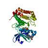
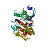
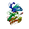
- PDB-5hu3: Drosophila CaMKII-D136N in complex with a phosphorylated fragment... -
+ Open data

- Basic information
| Entry | Database: PDB / ID: 5hu3 | ||||||
|---|---|---|---|---|---|---|---|
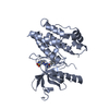
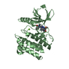
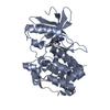
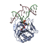
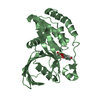
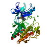
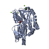
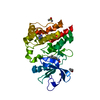
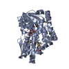
| Title | Drosophila CaMKII-D136N in complex with a phosphorylated fragment of the Eag potassium channel and Mg2+/ADP | ||||||
 Components Components |
| ||||||
 Keywords Keywords | TRANSFERASE / protein kinase / potassium channel / complex | ||||||
| Function / homology |  Function and homology information Function and homology informationperineurial glial growth / regulation of ovulation / Ca2+ pathway / Ion transport by P-type ATPases / HSF1-dependent transactivation / RAF activation / Ion homeostasis / Unblocking of NMDA receptors, glutamate binding and activation / positive regulation of synaptic transmission, dopaminergic / behavioral response to ether ...perineurial glial growth / regulation of ovulation / Ca2+ pathway / Ion transport by P-type ATPases / HSF1-dependent transactivation / RAF activation / Ion homeostasis / Unblocking of NMDA receptors, glutamate binding and activation / positive regulation of synaptic transmission, dopaminergic / behavioral response to ether / Voltage gated Potassium channels / regulation of synaptic assembly at neuromuscular junction / courtship behavior / male courtship behavior / Ca2+/calmodulin-dependent protein kinase / regulation of filopodium assembly / calcium/calmodulin-dependent protein kinase activity / negative regulation of cytokine production / presynaptic active zone / voltage-gated monoatomic cation channel activity / regulation of heart contraction / neuromuscular junction development / negative regulation of lipid storage / negative regulation of apoptotic signaling pathway / regulation of neuronal synaptic plasticity / voltage-gated potassium channel activity / long-term memory / regulation of protein localization to plasma membrane / voltage-gated potassium channel complex / potassium ion transmembrane transport / cellular response to starvation / learning / potassium ion transport / regulation of membrane potential / sensory perception of smell / chemical synaptic transmission / transmembrane transporter binding / learning or memory / calmodulin binding / postsynaptic membrane / neuron projection / postsynaptic density / protein serine kinase activity / axon / protein serine/threonine kinase activity / dendrite / ATP binding / plasma membrane / cytoplasm Similarity search - Function | ||||||
| Biological species |  | ||||||
| Method |  X-RAY DIFFRACTION / SYNCHROTRON / FOURIER SYNTHESIS / Resolution: 1.885 Å X-RAY DIFFRACTION / SYNCHROTRON / FOURIER SYNTHESIS / Resolution: 1.885 Å | ||||||
 Authors Authors | Castro-Rodrigues, A.F. / Morais-Cabral, J.H. | ||||||
 Citation Citation | Journal: J.Mol.Biol. / Year: 2018 Title: The Interaction between the Drosophila EAG Potassium Channel and the Protein Kinase CaMKII Involves an Extensive Interface at the Active Site of the Kinase. Authors: Castro-Rodrigues, A.F. / Zhao, Y. / Fonseca, F. / Gabant, G. / Cadene, M. / Robertson, G.A. / Morais-Cabral, J.H. | ||||||
| History |
|
- Structure visualization
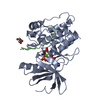
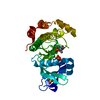
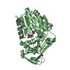
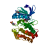
| Structure viewer | Molecule: MolmilJmol/JSmol |
|---|
- Downloads & links
-Download
| PDBx/mmCIF format | 5hu3.cif.gz | 136.3 KB | Display | PDBx/mmCIF format |
|---|---|---|---|---|
| PDB format | pdb5hu3.ent.gz | 103.7 KB | Display | PDB format |
| PDBx/mmJSON format | 5hu3.json.gz | Tree view | PDBx/mmJSON format | |
| Others |  Other downloads Other downloads |
-Validation report
| Arichive directory | https://data.pdbj.org/pub/pdb/validation_reports/hu/5hu3ftp://data.pdbj.org/pub/pdb/validation_reports/hu/5hu3 | HTTPS FTP |
|---|
-Related structure data


-Links
 PDBj
PDBj


- Assembly
| Deposited unit | 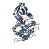
| ||||||||
|---|---|---|---|---|---|---|---|---|---|
| 1 |
| ||||||||
| Unit cell |
|
-Components
| #1: Protein | Mass: 32180.680 Da / Num. of mol.: 1 / Fragment: UNP Residues 1-283 / Mutation: D136N Source method: isolated from a genetically manipulated source Details: Constitutively active CaMKII kinase domain construct Source: (gene. exp.)  References: UniProt: Q00168, Ca2+/calmodulin-dependent protein kinase |
|---|---|
| #2: Protein | Mass: 5488.898 Da / Num. of mol.: 1 / Fragment: UNP Residues 768-820 Source method: isolated from a genetically manipulated source Details: Eag construct including the CaMKII-binding motif / Source: (gene. exp.) |
| #3: Chemical | ChemComp-ADP /   Mass: 427.201 Da / Num. of mol.: 1 / Source method: obtained synthetically / Formula: C10H15N5O10P2 / Comment: ADP, energy-carrying molecule*YM Mass: 427.201 Da / Num. of mol.: 1 / Source method: obtained synthetically / Formula: C10H15N5O10P2 / Comment: ADP, energy-carrying molecule*YM |
| #4: Chemical | ChemComp-MG /   Mass: 24.305 Da / Num. of mol.: 1 / Source method: obtained synthetically / Formula: Mg Mass: 24.305 Da / Num. of mol.: 1 / Source method: obtained synthetically / Formula: Mg |
| #5: Water | ChemComp-HOH /  Mass: 18.015 Da / Num. of mol.: 77 / Source method: isolated from a natural source / Formula: H2O Mass: 18.015 Da / Num. of mol.: 77 / Source method: isolated from a natural source / Formula: H2O |
| Has protein modification | Y |
-Experimental details
-Experiment
| Experiment | Method: X-RAY DIFFRACTION / Number of used crystals: 1 |
|---|
- Sample preparation
| Crystal | Density Matthews: 2.38 Å3/Da / Density % sol: 48.35 % |
|---|---|
| Crystal grow | Temperature: 293.15 K / Method: vapor diffusion, hanging drop / pH: 5 Details: 19% PEG 4000, 0.1 M sodium citrate pH 5.0, 0.2 M ammonium acetate, 5 mM magnesium chloride, 0.8 mM ADP PH range: 5.0 - 5.5 |
-Data collection
| Diffraction | Mean temperature: 100 K | ||||||||||||||||||||||||||||||||||||||||||||||||||||||||||||||||||||||||||||||||||||||||||||||||||||||||||||||||||||||||||||||||||||||||||||||||||||||||||||||||||||||||||||||||
|---|---|---|---|---|---|---|---|---|---|---|---|---|---|---|---|---|---|---|---|---|---|---|---|---|---|---|---|---|---|---|---|---|---|---|---|---|---|---|---|---|---|---|---|---|---|---|---|---|---|---|---|---|---|---|---|---|---|---|---|---|---|---|---|---|---|---|---|---|---|---|---|---|---|---|---|---|---|---|---|---|---|---|---|---|---|---|---|---|---|---|---|---|---|---|---|---|---|---|---|---|---|---|---|---|---|---|---|---|---|---|---|---|---|---|---|---|---|---|---|---|---|---|---|---|---|---|---|---|---|---|---|---|---|---|---|---|---|---|---|---|---|---|---|---|---|---|---|---|---|---|---|---|---|---|---|---|---|---|---|---|---|---|---|---|---|---|---|---|---|---|---|---|---|---|---|---|---|
| Diffraction source | Source: SYNCHROTRON / Site: ESRF  / Beamline: ID14-4 / Wavelength: 0.97625 Å / Beamline: ID14-4 / Wavelength: 0.97625 Å | ||||||||||||||||||||||||||||||||||||||||||||||||||||||||||||||||||||||||||||||||||||||||||||||||||||||||||||||||||||||||||||||||||||||||||||||||||||||||||||||||||||||||||||||||
| Detector | Type: ADSC QUANTUM 315r / Detector: CCD / Date: Jan 27, 2013 | ||||||||||||||||||||||||||||||||||||||||||||||||||||||||||||||||||||||||||||||||||||||||||||||||||||||||||||||||||||||||||||||||||||||||||||||||||||||||||||||||||||||||||||||||
| Radiation | Protocol: SINGLE WAVELENGTH / Monochromatic (M) / Laue (L): M / Scattering type: x-ray | ||||||||||||||||||||||||||||||||||||||||||||||||||||||||||||||||||||||||||||||||||||||||||||||||||||||||||||||||||||||||||||||||||||||||||||||||||||||||||||||||||||||||||||||||
| Radiation wavelength | Wavelength: 0.97625 Å / Relative weight: 1 | ||||||||||||||||||||||||||||||||||||||||||||||||||||||||||||||||||||||||||||||||||||||||||||||||||||||||||||||||||||||||||||||||||||||||||||||||||||||||||||||||||||||||||||||||
| Reflection | Resolution: 1.82→30.075 Å / Num. all: 26540 / Num. obs: 26540 / % possible obs: 97.9 % / Redundancy: 3.3 % / Biso Wilson estimate: 31.38 Å2 / Rpim(I) all: 0.022 / Rrim(I) all: 0.041 / Rsym value: 0.034 / Net I/av σ(I): 15.324 / Net I/σ(I): 19.3 / Num. measured all: 87981 | ||||||||||||||||||||||||||||||||||||||||||||||||||||||||||||||||||||||||||||||||||||||||||||||||||||||||||||||||||||||||||||||||||||||||||||||||||||||||||||||||||||||||||||||||
| Reflection shell | Diffraction-ID: 1 / Rejects: _
|
- Processing
| Software |
| ||||||||||||||||||||||||||||||||||||||||||||||||||||||||||||||||||||||||||||||||||||||||||||||||||||
|---|---|---|---|---|---|---|---|---|---|---|---|---|---|---|---|---|---|---|---|---|---|---|---|---|---|---|---|---|---|---|---|---|---|---|---|---|---|---|---|---|---|---|---|---|---|---|---|---|---|---|---|---|---|---|---|---|---|---|---|---|---|---|---|---|---|---|---|---|---|---|---|---|---|---|---|---|---|---|---|---|---|---|---|---|---|---|---|---|---|---|---|---|---|---|---|---|---|---|---|---|---|
| Refinement | Method to determine structure: FOURIER SYNTHESIS / Resolution: 1.885→30.075 Å / FOM work R set: 0.7364 / SU ML: 0.26 / Cross valid method: FREE R-VALUE / σ(F): 1.34 / Phase error: 31.82 / Stereochemistry target values: ML
| ||||||||||||||||||||||||||||||||||||||||||||||||||||||||||||||||||||||||||||||||||||||||||||||||||||
| Solvent computation | Shrinkage radii: 0.9 Å / VDW probe radii: 1.11 Å / Solvent model: FLAT BULK SOLVENT MODEL | ||||||||||||||||||||||||||||||||||||||||||||||||||||||||||||||||||||||||||||||||||||||||||||||||||||
| Displacement parameters | Biso max: 98.44 Å2 / Biso mean: 48.66 Å2 / Biso min: 16.94 Å2 | ||||||||||||||||||||||||||||||||||||||||||||||||||||||||||||||||||||||||||||||||||||||||||||||||||||
| Refinement step | Cycle: final / Resolution: 1.885→30.075 Å
| ||||||||||||||||||||||||||||||||||||||||||||||||||||||||||||||||||||||||||||||||||||||||||||||||||||
| Refine LS restraints |
| ||||||||||||||||||||||||||||||||||||||||||||||||||||||||||||||||||||||||||||||||||||||||||||||||||||
| LS refinement shell | Refine-ID: X-RAY DIFFRACTION / Total num. of bins used: 9
| ||||||||||||||||||||||||||||||||||||||||||||||||||||||||||||||||||||||||||||||||||||||||||||||||||||
| Refinement TLS params. | Method: refined / Refine-ID: X-RAY DIFFRACTION
| ||||||||||||||||||||||||||||||||||||||||||||||||||||||||||||||||||||||||||||||||||||||||||||||||||||
| Refinement TLS group |
|
