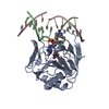
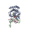
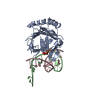
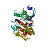
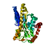
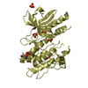
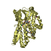
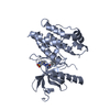
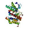
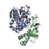
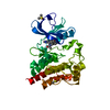
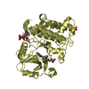
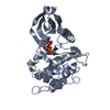
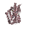
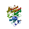
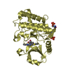
Alpha-ketoglutarate-dependentdioxygenasealkB / E.C.1.14.11.- / Alkylated DNA repair protein alkB
Mass: 22286.559 Da / Num. of mol.: 1 / Mutation: D135C Source method: isolated from a genetically manipulated source Source: (gene. exp.) Escherichia coli K12 (bacteria) / Species: Escherichia coli / Strain: K-12 / Gene: alkB, aidD / Plasmid: pET30 / Species (production host): Escherichia coli / Production host: Escherichia coli BL21(DE3) (bacteria) / Strain (production host): BL21(DE3) References: UniProt: P05050, Oxidoreductases; Acting on paired donors, with incorporation or reduction of molecular oxygen; With 2-oxoglutarate as one donor, and incorporation of one atom of oxygen into each donor
-
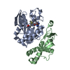
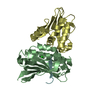
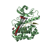
DNA chain , 2 types, 2 molecules BC
#2: DNA chain
DNA (5'-D(*DTP*DAP*DGP*DGP*DTP*DAP*DAP*DCP*DAP*DTP*DCP*DGP*DT)-3')
Mass: 4050.739 Da / Num. of mol.: 1 / Source method: obtained synthetically
#3: DNA chain
DNA (5'-D(*DAP*DAP*DCP*DGP*DAP*DTP*DAP*DTP*DTP*DAP*DCP*DCP*DT)-3')
Mass: 3934.599 Da / Num. of mol.: 1 / Source method: obtained synthetically
In the structure databanks used in Yorodumi, some data are registered as the other names, "COVID-19 virus" and "2019-nCoV". Here are the details of the virus and the list of structure data.
Jan 31, 2019. EMDB accession codes are about to change! (news from PDBe EMDB page)
EMDB accession codes are about to change! (news from PDBe EMDB page)
The allocation of 4 digits for EMDB accession codes will soon come to an end. Whilst these codes will remain in use, new EMDB accession codes will include an additional digit and will expand incrementally as the available range of codes is exhausted. The current 4-digit format prefixed with “EMD-” (i.e. EMD-XXXX) will advance to a 5-digit format (i.e. EMD-XXXXX), and so on. It is currently estimated that the 4-digit codes will be depleted around Spring 2019, at which point the 5-digit format will come into force.
The EM Navigator/Yorodumi systems omit the EMD- prefix.
Related info.:Q: What is EMD? / ID/Accession-code notation in Yorodumi/EM Navigator
Yorodumi is a browser for structure data from EMDB, PDB, SASBDB, etc.
This page is also the successor to EM Navigator detail page, and also detail information page/front-end page for Omokage search.
The word "yorodu" (or yorozu) is an old Japanese word meaning "ten thousand". "mi" (miru) is to see.
Related info.:EMDB / PDB / SASBDB / Comparison of 3 databanks / Yorodumi Search / Aug 31, 2016. New EM Navigator & Yorodumi / Yorodumi Papers / Jmol/JSmol / Function and homology information / Changes in new EM Navigator and Yorodumi
 Movie
Movie Controller

 Yorodumi
 Open data
 Basic information
 Components
 Keywords
 Function and homology information
 X-RAY DIFFRACTION /
  Authors
 Citation
 Structure visualization
 Downloads & links
 Other downloads
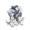


 PDBj
PDBj


 Assembly
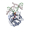


 Mass: 54.938 Da / Num. of mol.: 1 / Source method: obtained synthetically / Formula: Mn
 Mass: 146.098 Da / Num. of mol.: 1 / Source method: obtained synthetically / Formula: C5H6O5
 Sample preparation
 / Beamline: 14-BM-D / Wavelength: 0.97 Å
 Processing