 Movie
Movie Controller

[English] 日本語
 Yorodumi
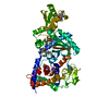
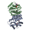
- PDB-2plr: Crystal structure of dTMP kinase (st1543) from Sulfolobus Tokodai... -
+ Open data

- Basic information
| Entry | Database: PDB / ID: 2plr | ||||||
|---|---|---|---|---|---|---|---|
| Title | Crystal structure of dTMP kinase (st1543) from Sulfolobus Tokodaii Strain7 | ||||||
 Components Components | Probable thymidylate kinase | ||||||
 Keywords Keywords | TRANSFERASE / TMP-binding / ATP-binding / Thymidylate Kinase / Structural Genomics / NPPSFA / National Project on Protein Structural and Functional Analyses / RIKEN Structural Genomics/Proteomics Initiative / RSGI | ||||||
| Function / homology |  Function and homology information Function and homology informationdTMP kinase / dUDP biosynthetic process / dTDP biosynthetic process / dTMP kinase activity / dTTP biosynthetic process / ATP binding / cytoplasm Similarity search - Function | ||||||
| Biological species |   Sulfolobus tokodaii (archaea) Sulfolobus tokodaii (archaea) | ||||||
| Method |  X-RAY DIFFRACTION / SYNCHROTRON / MOLECULAR REPLACEMENT / Resolution: 1.6 Å X-RAY DIFFRACTION / SYNCHROTRON / MOLECULAR REPLACEMENT / Resolution: 1.6 Å | ||||||
 Authors Authors | Kanaujia, S.P. / Jeyakanthan, J. / Rafi, Z.A. / Sekar, K. / Nakagawa, N. / Ebihara, A. / Kuramitsu, S. / Shinkai, A. / Shiro, Y. / Yokoyama, S. / RIKEN Structural Genomics/Proteomics Initiative (RSGI) | ||||||
 Citation Citation | Journal: To be Published Title: Crystal structure of dTMP kinase (st1543) from Sulfolobus Tokodaii Strain7 Authors: Kanaujia, S.P. / Jeyakanthan, J. / Rafi, Z.A. / Sekar, K. / Nakagawa, N. / Ebihara, A. / Kuramitsu, S. / Shinkai, A. / Shiro, Y. / Yokoyama, S. | ||||||
| History |
|
- Structure visualization
| Structure viewer | Molecule: MolmilJmol/JSmol |
|---|
- Downloads & links
-Download
| PDBx/mmCIF format | 2plr.cif.gz | 118.9 KB | Display | PDBx/mmCIF format |
|---|---|---|---|---|
| PDB format | pdb2plr.ent.gz | 89.4 KB | Display | PDB format |
| PDBx/mmJSON format | 2plr.json.gz | Tree view | PDBx/mmJSON format | |
| Others |  Other downloads Other downloads |
-Validation report
| Arichive directory | https://data.pdbj.org/pub/pdb/validation_reports/pl/2plrftp://data.pdbj.org/pub/pdb/validation_reports/pl/2plr | HTTPS FTP |
|---|
-Related structure data
| Related structure data |  2pbrS S: Starting model for refinement |
|---|---|
| Similar structure data | |
| Other databases |


-Links
 PDBj
PDBj


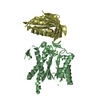
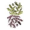
- Assembly
| Deposited unit | 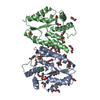
| ||||||||
|---|---|---|---|---|---|---|---|---|---|
| 1 |
| ||||||||
| Unit cell |
|
-Components
-Protein , 1 types, 2 molecules AB
| #1: Protein | Mass: 24725.273 Da / Num. of mol.: 2 Source method: isolated from a genetically manipulated source Source: (gene. exp.) Sulfolobus tokodaii (archaea) / Strain: 7 / Plasmid: pET21a / Production host:  |
|---|
-Non-polymers , 10 types, 550 molecules 


| #2: Chemical |  Mass: 35.453 Da / Num. of mol.: 2 / Source method: obtained synthetically / Formula: Cl Mass: 35.453 Da / Num. of mol.: 2 / Source method: obtained synthetically / Formula: Cl#3: Chemical | ChemComp-PO4 / |  Mass: 94.971 Da / Num. of mol.: 1 / Source method: obtained synthetically / Formula: PO4 Mass: 94.971 Da / Num. of mol.: 1 / Source method: obtained synthetically / Formula: PO4#4: Chemical |  Mass: 238.278 Da / Num. of mol.: 3 / Source method: obtained synthetically / Formula: C10H22O6 / Comment: precipitant*YM Mass: 238.278 Da / Num. of mol.: 3 / Source method: obtained synthetically / Formula: C10H22O6 / Comment: precipitant*YM#5: Chemical |  Mass: 150.173 Da / Num. of mol.: 3 / Source method: obtained synthetically / Formula: C6H14O4 Mass: 150.173 Da / Num. of mol.: 3 / Source method: obtained synthetically / Formula: C6H14O4#6: Chemical | ChemComp-EDO /  Mass: 62.068 Da / Num. of mol.: 6 / Source method: obtained synthetically / Formula: C2H6O2 Mass: 62.068 Da / Num. of mol.: 6 / Source method: obtained synthetically / Formula: C2H6O2#7: Chemical | ChemComp-DTT / |  Mass: 154.251 Da / Num. of mol.: 1 / Source method: obtained synthetically / Formula: C4H10O2S2 Mass: 154.251 Da / Num. of mol.: 1 / Source method: obtained synthetically / Formula: C4H10O2S2#8: Chemical | ChemComp-EPE / |  Mass: 238.305 Da / Num. of mol.: 1 / Source method: obtained synthetically / Formula: C8H18N2O4S / Comment: pH buffer*YM Mass: 238.305 Da / Num. of mol.: 1 / Source method: obtained synthetically / Formula: C8H18N2O4S / Comment: pH buffer*YM#9: Chemical | ChemComp-PEG /  Mass: 106.120 Da / Num. of mol.: 11 / Source method: obtained synthetically / Formula: C4H10O3 Mass: 106.120 Da / Num. of mol.: 11 / Source method: obtained synthetically / Formula: C4H10O3#10: Chemical | ChemComp-PG4 / |  Mass: 194.226 Da / Num. of mol.: 1 / Source method: obtained synthetically / Formula: C8H18O5 / Comment: precipitant*YM Mass: 194.226 Da / Num. of mol.: 1 / Source method: obtained synthetically / Formula: C8H18O5 / Comment: precipitant*YM#11: Water | ChemComp-HOH / | Mass: 18.015 Da / Num. of mol.: 521 / Source method: isolated from a natural source / Formula: H2O |
|---|
-Details
| Has protein modification | Y |
|---|
-Experimental details
-Experiment
| Experiment | Method: X-RAY DIFFRACTION / Number of used crystals: 1 |
|---|
- Sample preparation
| Crystal | Density Matthews: 2.16 Å3/Da / Density % sol: 43.1 % |
|---|---|
| Crystal grow | Temperature: 293 K / Method: vapor diffusion, hanging drop / pH: 7.5 Details: 50% PEG 200, 100mM Hepes, pH 7.5, VAPOR DIFFUSION, HANGING DROP, temperature 293K |
-Data collection
| Diffraction | Mean temperature: 100 K | ||||||||||||
|---|---|---|---|---|---|---|---|---|---|---|---|---|---|
| Diffraction source | Source: SYNCHROTRON / Site: SPring-8  / Beamline: BL26B2 / Wavelength: 0.9789, 0.9793, 0.9000 / Beamline: BL26B2 / Wavelength: 0.9789, 0.9793, 0.9000 | ||||||||||||
| Detector | Type: RIGAKU RAXIS V / Detector: IMAGE PLATE / Date: Jun 26, 2006 / Details: RH Coated Bent-Cyrindrical MIRROR | ||||||||||||
| Radiation | Monochromator: SI-1 1 1 Double Crystal Monochromator / Protocol: MAD / Monochromatic (M) / Laue (L): M / Scattering type: x-ray | ||||||||||||
| Radiation wavelength |
| ||||||||||||
| Reflection | Resolution: 1.6→50 Å / Num. all: 57129 / Num. obs: 57129 / % possible obs: 99.4 % / Biso Wilson estimate: 18.3 Å2 / Rmerge(I) obs: 0.041 / Rsym value: 0.043 | ||||||||||||
| Reflection shell | Resolution: 1.6→1.66 Å / Rmerge(I) obs: 0.209 / Num. unique all: 5414 / Rsym value: 0.23 / % possible all: 95.5 |
- Processing
| Software |
| ||||||||||||||||||||
|---|---|---|---|---|---|---|---|---|---|---|---|---|---|---|---|---|---|---|---|---|---|
| Refinement | Method to determine structure: MOLECULAR REPLACEMENT Starting model: PDB ENTRY 2PBR Resolution: 1.6→46.59 Å / Rfactor Rfree error: 0.003 / Data cutoff high absF: 7231113.62 / Data cutoff low absF: 0 / Isotropic thermal model: RESTRAINED / Cross valid method: THROUGHOUT / σ(F): 0 / Stereochemistry target values: Engh & Huber
| ||||||||||||||||||||
| Solvent computation | Solvent model: FLAT MODEL / Bsol: 64.913 Å2 / ksol: 0.374268 e/Å3 | ||||||||||||||||||||
| Displacement parameters | Biso mean: 24.9 Å2
| ||||||||||||||||||||
| Refine analyze |
| ||||||||||||||||||||
| Refinement step | Cycle: LAST / Resolution: 1.6→46.59 Å
| ||||||||||||||||||||
| Refine LS restraints |
| ||||||||||||||||||||
| LS refinement shell | Resolution: 1.6→1.67 Å / Rfactor Rfree error: 0.011 / Total num. of bins used: 8
| ||||||||||||||||||||
| Xplor file |
|
