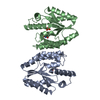
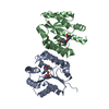
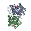
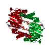
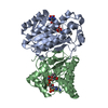
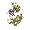
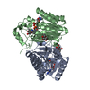
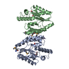
Entry Database : PDB / ID : 4mqbTitle Crystal structure of thymidylate kinase from Staphylococcus aureus in complex with 2-(N-morpholino)ethanesulfonic acid Thymidylate kinase Keywords / / / / / / / / Function / homology Function Domain/homology Component
/ / / / / / / / / / / / / / / Biological species Staphylococcus aureus (bacteria)Method / / / Resolution : 1.55 Å Authors Filippova, E.V. / Minasov, G. / Shuvalova, L. / Kiryukhina, O. / Jedrzejczak, R. / Babnigg, G. / Rubin, E. / Sacchettini, J. / Joachimiak, A. / Anderson, W.F. ...Filippova, E.V. / Minasov, G. / Shuvalova, L. / Kiryukhina, O. / Jedrzejczak, R. / Babnigg, G. / Rubin, E. / Sacchettini, J. / Joachimiak, A. / Anderson, W.F. / Midwest Center for Structural Genomics (MCSG) / Structures of Mtb Proteins Conferring Susceptibility to Known Mtb Inhibitors (MTBI) Journal : To be Published Title : Crystal structure of thymidylate kinase from Staphylococcus aureus in complex with 2-(N-morpholino)ethanesulfonic acidAuthors : Filippova, E.V. / Minasov, G. / Shuvalova, L. / Kiryukhina, O. / Jedrzejczak, R. / Babnigg, G. / Rubin, E. / Sacchettini, J. / Joachimiak, A. / Anderson, W.F. History Deposition Sep 16, 2013 Deposition site / Processing site Revision 1.0 Oct 23, 2013 Provider / Type Revision 1.1 Nov 15, 2017 Group / Category / Item Revision 1.2 Sep 20, 2023 Group Data collection / Database references ... Data collection / Database references / Derived calculations / Refinement description Category chem_comp_atom / chem_comp_bond ... chem_comp_atom / chem_comp_bond / database_2 / pdbx_initial_refinement_model / struct_conn / struct_ref_seq_dif / struct_site Item _database_2.pdbx_DOI / _database_2.pdbx_database_accession ... _database_2.pdbx_DOI / _database_2.pdbx_database_accession / _struct_conn.pdbx_leaving_atom_flag / _struct_ref_seq_dif.details / _struct_site.pdbx_auth_asym_id / _struct_site.pdbx_auth_comp_id / _struct_site.pdbx_auth_seq_id Revision 1.3 Dec 6, 2023 Group / Category / chem_comp_bond / Item / _chem_comp_bond.atom_id_2Revision 1.4 Oct 9, 2024 Group / Category / pdbx_modification_feature
Show all Show less
 Movie
Movie Controller

 Yorodumi
 Open data
 Basic information
 Components
 Keywords
 Function and homology information
 Staphylococcus aureus (bacteria)
 X-RAY DIFFRACTION /
  Authors
 Citation
 Structure visualization
 Downloads & links
 Other downloads
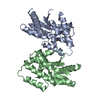

 PDBj
PDBj Assembly
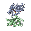

 Mass: 195.237 Da / Num. of mol.: 1 / Source method: obtained synthetically / Formula: C6H13NO4S / Comment: pH buffer*YM
 Mass: 194.226 Da / Num. of mol.: 2 / Source method: obtained synthetically / Formula: C8H18O5 / Comment: precipitant*YM
 Mass: 18.015 Da / Num. of mol.: 329 / Source method: isolated from a natural source / Formula: H2O
 Sample preparation
 / Beamline: 21-ID-G / Wavelength: 0.97856 Å
 Processing