[English] 日本語
Yorodumi
- PDB-6uyk: Dark-operative protochlorophyllide oxidoreductase in the nucleoti... -

+
Open data


ID or keywords:

Loading...

-
Basic information

Entry
Database: PDB / ID: 6uyk
Title
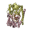
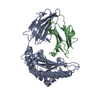
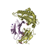
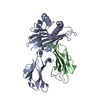
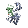
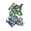
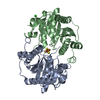
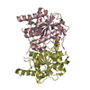
Dark-operative protochlorophyllide oxidoreductase in the nucleotide-free form.
ComponentsLight-independent protochlorophyllide reductase iron-sulfur ATP-binding protein
KeywordsOXIDOREDUCTASE / Electron Transfer / Nitrogenase / DPOR / Photosynthesis / Iron-Sulfur cluster
Function / homology
Function and homology information


ferredoxin:protochlorophyllide reductase (ATP-dependent) / photosynthesis, dark reaction / light-independent bacteriochlorophyll biosynthetic process / oxidoreductase activity, acting on the CH-CH group of donors, iron-sulfur protein as acceptor / oxidoreductase activity, acting on iron-sulfur proteins as donors / 4 iron, 4 sulfur cluster binding / ATP binding / metal ion binding
Similarity search - Function
Light-independent protochlorophyllide reductase, iron-sulphur ATP-binding protein / NifH/frxC family / NifH/chlL conserved site / 4Fe-4S iron sulfur cluster binding proteins, NifH/frxC family / NifH/frxC family signature 2. / NifH/frxC family signature 1. / NIFH_FRXC family profile. / P-loop containing nucleoside triphosphate hydrolase
Similarity search - Domain/homology
IRON/SULFUR CLUSTER / Light-independent protochlorophyllide reductase iron-sulfur ATP-binding protein
Similarity search - Component
Biological speciesRhodobacter sphaeroides (bacteria)
MethodX-RAY DIFFRACTION / SYNCHROTRON / MOLECULAR REPLACEMENT / Resolution: 2.6 Å
AuthorsBacik, J.P. / Imran, S.M.S. / Watkins, M.B. / Corless, E. / Antony, E. / Ando, N.
Funding support United States, 1items
OrganizationGrant numberCountry
Department of Energy (DOE, United States)DE-SC0017866 United States
CitationJournal: J.Biol.Chem. / Year: 2020
Title: The flexible N-terminus of BchL autoinhibits activity through interaction with its [4Fe-4S] cluster and released upon ATP binding.
Authors: Corless, E.I. / Saad Imran, S.M. / Watkins, M.B. / Bacik, J.P. / Mattice, J.R. / Patterson, A. / Danyal, K. / Soffe, M. / Kitelinger, R. / Seefeldt, L.C. / Origanti, S. / Bennett, B. / ...Authors: Corless, E.I. / Saad Imran, S.M. / Watkins, M.B. / Bacik, J.P. / Mattice, J.R. / Patterson, A. / Danyal, K. / Soffe, M. / Kitelinger, R. / Seefeldt, L.C. / Origanti, S. / Bennett, B. / Bothner, B. / Ando, N. / Antony, E.
History
DepositionNov 13, 2019Deposition site: RCSB / Processing site: RCSB
Revision 1.0Dec 2, 2020Provider: repository / Type: Initial release
Revision 1.1Apr 28, 2021Group: Database references / Category: citation / citation_author
Item: _citation.page_first / _citation.page_last / _citation_author.identifier_ORCID
Revision 1.2Jul 14, 2021Group: Database references / Category: citation / citation_author
Item: _citation.journal_volume / _citation.title / _citation_author.name
Revision 1.3Oct 11, 2023Group: Data collection / Database references / Refinement description
Category: chem_comp_atom / chem_comp_bond ...chem_comp_atom / chem_comp_bond / database_2 / pdbx_initial_refinement_model
Item: _database_2.pdbx_DOI / _database_2.pdbx_database_accession

-
Structure visualization

Structure viewerMolecule:
MolmilJmol/JSmol

Downloads & links

-
Assembly

Deposited unit
A: Light-independent protochlorophyllide reductase iron-sulfur ATP-binding protein
B: Light-independent protochlorophyllide reductase iron-sulfur ATP-binding protein
C: Light-independent protochlorophyllide reductase iron-sulfur ATP-binding protein
D: Light-independent protochlorophyllide reductase iron-sulfur ATP-binding protein
hetero molecules


Theoretical massNumber of molelcules
Total (without water)140,1768
Polymers139,4014
Non-polymers7744
Water181
1
A: Light-independent protochlorophyllide reductase iron-sulfur ATP-binding protein
B: Light-independent protochlorophyllide reductase iron-sulfur ATP-binding protein
hetero molecules


Theoretical massNumber of molelcules
Total (without water)70,0884
Polymers69,7012
Non-polymers3872
Water181
TypeNameSymmetry operationNumber
identity operation1_555x,y,z1
Buried area2730 Å2
ΔGint-39 kcal/mol
Surface area20600 Å2
MethodPISA
2
C: Light-independent protochlorophyllide reductase iron-sulfur ATP-binding protein
D: Light-independent protochlorophyllide reductase iron-sulfur ATP-binding protein
hetero molecules


Theoretical massNumber of molelcules
Total (without water)70,0884
Polymers69,7012
Non-polymers3872
Water0
TypeNameSymmetry operationNumber
identity operation1_555x,y,z1
Buried area3260 Å2
ΔGint-49 kcal/mol
Surface area20620 Å2
MethodPISA
Unit cell
Length a, b, c (Å)92.513, 100.918, 117.789
Angle α, β, γ (deg.)90.000, 99.270, 90.000
Int Tables number5
Space group name H-MI121
Space group name HallC2y(x,y,-x+z)
Symmetry operation#1: x,y,z
#2: -x,y,-z
#3: x+1/2,y+1/2,z+1/2
#4: -x+1/2,y+1/2,-z+1/2

-
Components

#1: Protein
Light-independent protochlorophyllide reductase iron-sulfur ATP-binding protein / LI-POR subunit L


Mass: 34850.340 Da / Num. of mol.: 4
Source method: isolated from a genetically manipulated source
Source: (gene. exp.) Rhodobacter sphaeroides (bacteria) / Gene: bchL, RHOS4_18930, RSP_0288 / Production host: Escherichia coli (E. coli)
References: UniProt: Q9RFD6, ferredoxin:protochlorophyllide reductase (ATP-dependent)
#2: Chemical ChemComp-SF4 / IRON/SULFUR CLUSTER / Iron–sulfur cluster


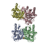
Mass: 351.640 Da / Num. of mol.: 2 / Source method: obtained synthetically / Formula: Fe4S4 / Feature type: SUBJECT OF INVESTIGATION
#3: Chemical ChemComp-CL / CHLORIDE ION / Chloride


Mass: 35.453 Da / Num. of mol.: 2 / Source method: obtained synthetically / Formula: Cl
#4: Water ChemComp-HOH / water / Water


Mass: 18.015 Da / Num. of mol.: 1 / Source method: isolated from a natural source / Formula: H2O
Has ligand of interestY

-
Experimental details

-
Experiment

ExperimentMethod: X-RAY DIFFRACTION / Number of used crystals: 1

-
Sample preparation

CrystalDensity Matthews: 1.95 Å3/Da / Density % sol: 36.81 %
Crystal growTemperature: 288 K / Method: vapor diffusion, sitting drop
Details: 1 ul BchL protein in 100 mM HEPES pH 7.5, 150 mM NaCl, 10% (v/v) glycerol was mixed with 2 ul well solution containing 0.6 M sodium chloride, 0.1 M MES:NaOH pH 6.5, 20% (w/v) PEG 4000. Prior ...Details: 1 ul BchL protein in 100 mM HEPES pH 7.5, 150 mM NaCl, 10% (v/v) glycerol was mixed with 2 ul well solution containing 0.6 M sodium chloride, 0.1 M MES:NaOH pH 6.5, 20% (w/v) PEG 4000. Prior to freezing, the well solution was mixed in an equal volume of cryoprotectant solution with a final concentration of 9% (w/v) sucrose, 2% (w/v) glucose, 8% (v/v) glycerol, and 8% (v/v) ethylene glycol. Crystals were soaked for a few seconds in the cryoprotectant before being frozen in liquid nitrogen

-
Data collection

DiffractionMean temperature: 100 K / Serial crystal experiment: N
Diffraction sourceSource: SYNCHROTRON / Site: NSLS-II / Beamline: 17-ID-1 / Wavelength: 1.008 Å
DetectorType: DECTRIS EIGER X 9M / Detector: PIXEL / Date: Apr 9, 2018
RadiationProtocol: SINGLE WAVELENGTH / Monochromatic (M) / Laue (L): M / Scattering type: x-ray
Radiation wavelengthWavelength: 1.008 Å / Relative weight: 1
ReflectionResolution: 2.6→42.2 Å / Num. obs: 32733 / % possible obs: 99.5 % / Redundancy: 9.3 % / Biso Wilson estimate: 80.66 Å2 / CC1/2: 0.99 / Rmerge(I) obs: 0.173 / Rpim(I) all: 0.057 / Net I/σ(I): 9.2
Reflection shellResolution: 2.6→2.69 Å / Redundancy: 5.1 % / Rmerge(I) obs: 0.908 / Mean I/σ(I) obs: 1.5 / Num. unique obs: 3125 / CC1/2: 0.573 / Rpim(I) all: 0.413 / % possible all: 95.3

-
Processing

Software
NameVersionClassification
PHENIX1.13_2998refinement
HKL-2000data reduction
HKL-2000data scaling
PHASERphasing
RefinementMethod to determine structure: MOLECULAR REPLACEMENT
Starting model: 3FWY
Resolution: 2.6→41.71 Å / SU ML: 0.4791 / Cross valid method: FREE R-VALUE / σ(F): 1.34 / Phase error: 34.6425
RfactorNum. reflection% reflection
Rfree0.2772 2873 8.78 %
Rwork0.2314 --
obs0.2354 32732 99.41 %
Solvent computationShrinkage radii: 0.9 Å / VDW probe radii: 1.11 Å
Displacement parametersBiso mean: 85.6 Å2
Refinement stepCycle: LAST / Resolution: 2.6→41.71 Å
ProteinNucleic acidLigandSolventTotal
Num. atoms7648 0 18 1 7667
Refine LS restraints
Refine-IDTypeDev idealNumber
X-RAY DIFFRACTIONf_bond_d0.00197814
X-RAY DIFFRACTIONf_angle_d0.39310651
X-RAY DIFFRACTIONf_chiral_restr0.03981255
X-RAY DIFFRACTIONf_plane_restr0.00341396
X-RAY DIFFRACTIONf_dihedral_angle_d14.00582690
LS refinement shell
Resolution (Å)Rfactor RfreeNum. reflection RfreeRfactor RworkNum. reflection RworkRefine-ID% reflection obs (%)
2.6-2.640.44751290.35511325X-RAY DIFFRACTION91.1
2.64-2.690.38341310.34521356X-RAY DIFFRACTION97.19
2.69-2.740.41921360.33541418X-RAY DIFFRACTION99.49
2.74-2.790.3721360.32111417X-RAY DIFFRACTION100
2.79-2.850.33841250.31441436X-RAY DIFFRACTION100
2.85-2.910.34091250.30651414X-RAY DIFFRACTION100
2.91-2.980.34151470.29791449X-RAY DIFFRACTION100
2.98-3.050.3581340.29571425X-RAY DIFFRACTION100
3.05-3.130.40471340.30861414X-RAY DIFFRACTION100
3.13-3.230.36431400.291398X-RAY DIFFRACTION100
3.23-3.330.31431410.28731433X-RAY DIFFRACTION100
3.33-3.450.3451420.26451428X-RAY DIFFRACTION100
3.45-3.590.34771360.24641432X-RAY DIFFRACTION100
3.59-3.750.31461380.25331410X-RAY DIFFRACTION100
3.75-3.950.28421430.24331432X-RAY DIFFRACTION100
3.95-4.20.27881360.22261436X-RAY DIFFRACTION100
4.2-4.520.25121400.20551450X-RAY DIFFRACTION100
4.52-4.970.24211380.19941413X-RAY DIFFRACTION100
4.97-5.690.27261400.21171457X-RAY DIFFRACTION100
5.69-7.160.26481390.22661440X-RAY DIFFRACTION100
7.16-41.710.18931430.17911476X-RAY DIFFRACTION99.94

+
About Yorodumi

-
News

-
Feb 9, 2022. New format data for meta-information of EMDB entries

New format data for meta-information of EMDB entries

  • Version 3 of the EMDB header file is now the official format.
  • The previous official version 1.9 will be removed from the archive.

Related info.:EMDB header

External links:wwPDB to switch to version 3 of the EMDB data model

-
Aug 12, 2020. Covid-19 info

Covid-19 info

URL: https://pdbj.org/emnavi/covid19.php

New page: Covid-19 featured information page in EM Navigator.

Related info.:Covid-19 info / Mar 5, 2020. Novel coronavirus structure data

+
Mar 5, 2020. Novel coronavirus structure data

Novel coronavirus structure data

Related info.:Yorodumi Speices / Aug 12, 2020. Covid-19 info

External links:COVID-19 featured content - PDBj / Molecule of the Month (242):Coronavirus Proteases

+
Jan 31, 2019. EMDB accession codes are about to change! (news from PDBe EMDB page)

EMDB accession codes are about to change! (news from PDBe EMDB page)

  • The allocation of 4 digits for EMDB accession codes will soon come to an end. Whilst these codes will remain in use, new EMDB accession codes will include an additional digit and will expand incrementally as the available range of codes is exhausted. The current 4-digit format prefixed with “EMD-” (i.e. EMD-XXXX) will advance to a 5-digit format (i.e. EMD-XXXXX), and so on. It is currently estimated that the 4-digit codes will be depleted around Spring 2019, at which point the 5-digit format will come into force.
  • The EM Navigator/Yorodumi systems omit the EMD- prefix.

Related info.:Q: What is EMD? / ID/Accession-code notation in Yorodumi/EM Navigator

External links:EMDB Accession Codes are Changing Soon! / Contact to PDBj

+
Jul 12, 2017. Major update of PDB

Major update of PDB

  • wwPDB released updated PDB data conforming to the new PDBx/mmCIF dictionary.
  • This is a major update changing the version number from 4 to 5, and with Remediation, in which all the entries are updated.
  • In this update, many items about electron microscopy experimental information are reorganized (e.g. em_software).
  • Now, EM Navigator and Yorodumi are based on the updated data.

External links:wwPDB Remediation / Enriched Model Files Conforming to OneDep Data Standards Now Available in the PDB FTP Archive

-
Yorodumi

Thousand views of thousand structures

  • Yorodumi is a browser for structure data from EMDB, PDB, SASBDB, etc.
  • This page is also the successor to EM Navigator detail page, and also detail information page/front-end page for Omokage search.
  • The word "yorodu" (or yorozu) is an old Japanese word meaning "ten thousand". "mi" (miru) is to see.

Related info.:EMDB / PDB / SASBDB / Comparison of 3 databanks / Yorodumi Search / Aug 31, 2016. New EM Navigator & Yorodumi / Yorodumi Papers / Jmol/JSmol / Function and homology information / Changes in new EM Navigator and Yorodumi

Read more