 Movie
Movie Controller

[English] 日本語
 Yorodumi
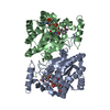
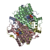
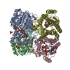
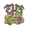
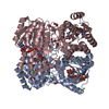
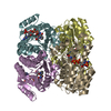
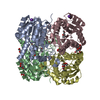
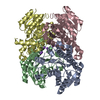
- PDB-1g0n: STRUCTURE OF TRIHYDROXYNAPHTHALENE REDUCTASE IN COMPLEX WITH NADP... -
+ Open data

- Basic information
| Entry | Database: PDB / ID: 1g0n | ||||||
|---|---|---|---|---|---|---|---|
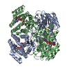
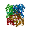
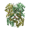
| Title | STRUCTURE OF TRIHYDROXYNAPHTHALENE REDUCTASE IN COMPLEX WITH NADPH AND 4,5,6,7-TETRACHLORO-PHTHALIDE | ||||||
 Components Components | TRIHYDROXYNAPHTHALENE REDUCTASE | ||||||
 Keywords Keywords | OXIDOREDUCTASE / Protein-NADPH-active site inhibitor complex / diuncleotide binding fold / short chain dehydrogenase | ||||||
| Function / homology |  Function and homology information Function and homology informationtetrahydroxynaphthalene reductase / tetrahydroxynaphthalene reductase activity / melanin biosynthetic process Similarity search - Function | ||||||
| Biological species |  Magnaporthe grisea (fungus) Magnaporthe grisea (fungus) | ||||||
| Method |  X-RAY DIFFRACTION / Resolution: 2 Å X-RAY DIFFRACTION / Resolution: 2 Å | ||||||
 Authors Authors | Liao, D. / Basarab, G.S. / Gatenby, A.A. / Valent, B. / Jordan, D.B. | ||||||
 Citation Citation | Journal: Structure / Year: 2001 Title: Structures of trihydroxynaphthalene reductase-fungicide complexes: implications for structure-based design and catalysis. Authors: Liao, D. / Basarab, G.S. / Gatenby, A.A. / Valent, B. / Jordan, D.B. | ||||||
| History |
|
- Structure visualization
| Structure viewer | Molecule: MolmilJmol/JSmol |
|---|
- Downloads & links
-Download
| PDBx/mmCIF format | 1g0n.cif.gz | 121.5 KB | Display | PDBx/mmCIF format |
|---|---|---|---|---|
| PDB format | pdb1g0n.ent.gz | 94.3 KB | Display | PDB format |
| PDBx/mmJSON format | 1g0n.json.gz | Tree view | PDBx/mmJSON format | |
| Others |  Other downloads Other downloads |
-Validation report
| Arichive directory | https://data.pdbj.org/pub/pdb/validation_reports/g0/1g0nftp://data.pdbj.org/pub/pdb/validation_reports/g0/1g0n | HTTPS FTP |
|---|
-Related structure data


-Links
 PDBj
PDBj


- Assembly
| Deposited unit | 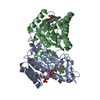
| ||||||||
|---|---|---|---|---|---|---|---|---|---|
| 1 | 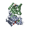
| ||||||||
| Unit cell |
| ||||||||
| Details | The biological assembly is a homo tetramer. The two molecules in the asymmetric unit represent half of the biological assembly. The tetramer has a 222 symmetry / The following transformation should be applied to both chain A and chain B to generate the tetramer. Rotation matrix: -0.500 -0.866 0.00 -0.866 0.500 0.00 0.00 0.00 -1.00 Translation vector: 214.21, 123.66, 24.31 |
-Components
| #1: Protein | Mass: 30177.682 Da / Num. of mol.: 2 / Mutation: S241V, A242Q, H247R Source method: isolated from a genetically manipulated source Source: (gene. exp.) Magnaporthe grisea (fungus) / Plasmid: PTHNR2 / Production host:  References: UniProt: Q12634, tetrahydroxynaphthalene reductase #2: Chemical |   Mass: 745.421 Da / Num. of mol.: 2 / Source method: obtained synthetically / Formula: C21H30N7O17P3 Mass: 745.421 Da / Num. of mol.: 2 / Source method: obtained synthetically / Formula: C21H30N7O17P3#3: Chemical | ChemComp-PHH / | 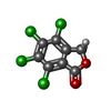  Mass: 271.912 Da / Num. of mol.: 1 / Source method: obtained synthetically / Formula: C8H2Cl4O2 Mass: 271.912 Da / Num. of mol.: 1 / Source method: obtained synthetically / Formula: C8H2Cl4O2#4: Water | ChemComp-HOH / |  Mass: 18.015 Da / Num. of mol.: 386 / Source method: isolated from a natural source / Formula: H2O Mass: 18.015 Da / Num. of mol.: 386 / Source method: isolated from a natural source / Formula: H2O |
|---|
-Experimental details
-Experiment
| Experiment | Method: X-RAY DIFFRACTION / Number of used crystals: 1 |
|---|
- Sample preparation
| Crystal | Density Matthews: 3.43 Å3/Da / Density % sol: 64.15 % | ||||||||||||||||||||||||||||||||||||||||
|---|---|---|---|---|---|---|---|---|---|---|---|---|---|---|---|---|---|---|---|---|---|---|---|---|---|---|---|---|---|---|---|---|---|---|---|---|---|---|---|---|---|
| Crystal grow | Temperature: 274 K / Method: vapor diffusion, hanging drop / pH: 7 Details: PEG 6K, glycerol, Hepes, pH 7.0, VAPOR DIFFUSION, HANGING DROP, temperature 274K | ||||||||||||||||||||||||||||||||||||||||
| Crystal grow | *PLUS Temperature: 4 ℃ | ||||||||||||||||||||||||||||||||||||||||
| Components of the solutions | *PLUS
|
-Data collection
| Diffraction | Mean temperature: 102 K |
|---|---|
| Diffraction source | Source: ROTATING ANODE / Type: RIGAKU RU200 / Wavelength: 1.54 |
| Detector | Type: RIGAKU RAXIS IV / Detector: IMAGE PLATE / Date: Aug 26, 1997 |
| Radiation | Protocol: SINGLE WAVELENGTH / Monochromatic (M) / Laue (L): M / Scattering type: x-ray |
| Radiation wavelength | Wavelength: 1.54 Å / Relative weight: 1 |
| Reflection | Resolution: 2→30 Å / Num. all: 54812 / Num. obs: 208682 / % possible obs: 96.5 % / Observed criterion σ(I): 0 / Redundancy: 3.8 % / Rmerge(I) obs: 0.081 / Net I/σ(I): 15.1 |
| Reflection shell | Resolution: 2→2.03 Å / Redundancy: 2.7 % / Rmerge(I) obs: 0.351 / Num. unique all: 2189 / % possible all: 76.4 |
| Reflection | *PLUS Num. obs: 54812 / Num. measured all: 208682 |
| Reflection shell | *PLUS % possible obs: 76.4 % |
- Processing
| Software |
| |||||||||||||||
|---|---|---|---|---|---|---|---|---|---|---|---|---|---|---|---|---|
| Refinement | Resolution: 2→8 Å / σ(F): 2 / Stereochemistry target values: Engh & Huber Details: Only one monomer of the dimer in the asymmetric unit has the electron density for the inhibitor.
| |||||||||||||||
| Refinement step | Cycle: LAST / Resolution: 2→8 Å
| |||||||||||||||
| Refine LS restraints |
| |||||||||||||||
| Software | *PLUS Name: X-PLOR / Version: 3.1 / Classification: refinement | |||||||||||||||
| Refinement | *PLUS Highest resolution: 2 Å / Lowest resolution: 8 Å / σ(F): 2 / % reflection Rfree: 10 % / Rfactor obs: 0.195 | |||||||||||||||
| Solvent computation | *PLUS | |||||||||||||||
| Displacement parameters | *PLUS |
