 Movie
Movie Controller

[English] 日本語
 Yorodumi
- PDB-2pbr: Crystal structure of thymidylate kinase (aq_969) from Aquifex Aeo... -
+ Open data

- Basic information
| Entry | Database: PDB / ID: 2pbr | ||||||
|---|---|---|---|---|---|---|---|
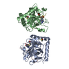
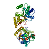
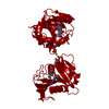
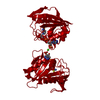
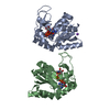
| Title | Crystal structure of thymidylate kinase (aq_969) from Aquifex Aeolicus VF5 | ||||||
 Components Components | Thymidylate kinase | ||||||
 Keywords Keywords | TRANSFERASE / KINASE / NUCLEOTIDE BIOSYNTHESIS / TMP-BINDING / ATP-BINDING / Structural Genomics / NPPSFA / National Project on Protein Structural and Functional Analyses / RIKEN Structural Genomics/Proteomics Initiative / RSGI | ||||||
| Function / homology |  Function and homology information Function and homology informationdTMP kinase / dUDP biosynthetic process / dTDP biosynthetic process / dTMP kinase activity / dTTP biosynthetic process / ATP binding / cytoplasm / cytosol Similarity search - Function | ||||||
| Biological species |   Aquifex aeolicus (bacteria) Aquifex aeolicus (bacteria) | ||||||
| Method |  X-RAY DIFFRACTION / SYNCHROTRON / MOLECULAR REPLACEMENT / Resolution: 1.96 Å X-RAY DIFFRACTION / SYNCHROTRON / MOLECULAR REPLACEMENT / Resolution: 1.96 Å | ||||||
 Authors Authors | Jeyakanthan, J. / Kanaujia, S.P. / Vasuki Ranjani, C. / Sekar, K. / Nakagawa, N. / Ebihara, A. / Kuramitsu, S. / Shinkai, A. / Shiro, Y. / Yokoyama, S. / RIKEN Structural Genomics/Proteomics Initiative (RSGI) | ||||||
 Citation Citation | Journal: To be Published Title: Crystal structure of thymidylate kinase (aq_969) from Aquifex Aeolicus VF5 Authors: Jeyakanthan, J. / Kanaujia, S.P. / Vasuki Ranjani, C. / Sekar, K. / Nakagawa, N. / Ebihara, A. / Kuramitsu, S. / Shinkai, A. / Shiro, Y. / Yokoyama, S. | ||||||
| History |
|
- Structure visualization
| Structure viewer | Molecule: MolmilJmol/JSmol |
|---|
- Downloads & links
-Download
| PDBx/mmCIF format | 2pbr.cif.gz | 95.6 KB | Display | PDBx/mmCIF format |
|---|---|---|---|---|
| PDB format | pdb2pbr.ent.gz | 72.8 KB | Display | PDB format |
| PDBx/mmJSON format | 2pbr.json.gz | Tree view | PDBx/mmJSON format | |
| Others |  Other downloads Other downloads |
-Validation report
| Arichive directory | https://data.pdbj.org/pub/pdb/validation_reports/pb/2pbrftp://data.pdbj.org/pub/pdb/validation_reports/pb/2pbr | HTTPS FTP |
|---|
-Related structure data
| Related structure data | 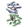 2ccjS S: Starting model for refinement |
|---|---|
| Similar structure data | |
| Other databases |


-Links
 PDBj
PDBj- Assembly
| Deposited unit | 
| ||||||||
|---|---|---|---|---|---|---|---|---|---|
| 1 |
| ||||||||
| 2 | 
| ||||||||
| Unit cell |
|
-Components
| #1: Protein | Mass: 22402.961 Da / Num. of mol.: 2 Source method: isolated from a genetically manipulated source Source: (gene. exp.) Aquifex aeolicus (bacteria) / Strain: VF5 / Plasmid: PET21A / Production host: #2: Chemical |   Mass: 96.063 Da / Num. of mol.: 2 / Source method: obtained synthetically / Formula: SO4 Mass: 96.063 Da / Num. of mol.: 2 / Source method: obtained synthetically / Formula: SO4#3: Water | ChemComp-HOH / |  Mass: 18.015 Da / Num. of mol.: 320 / Source method: isolated from a natural source / Formula: H2O Mass: 18.015 Da / Num. of mol.: 320 / Source method: isolated from a natural source / Formula: H2O |
|---|
-Experimental details
-Experiment
| Experiment | Method: X-RAY DIFFRACTION / Number of used crystals: 1 |
|---|
- Sample preparation
| Crystal | Density Matthews: 2.35 Å3/Da / Density % sol: 47.7 % |
|---|---|
| Crystal grow | Temperature: 293 K / Method: vapor diffusion, sitting drop / pH: 6.6 Details: 0.1 M ADA, 0.05 M Lithium Sulfate, 12% PEG 4000, 2% iso-propanol, 3% D(+)-sucrose as additive, pH 6.6, VAPOR DIFFUSION, SITTING DROP, temperature 293K |
-Data collection
| Diffraction | Mean temperature: 100 K |
|---|---|
| Diffraction source | Source: SYNCHROTRON / Site: SPring-8  / Beamline: BL26B2 / Wavelength: 1 Å / Beamline: BL26B2 / Wavelength: 1 Å |
| Detector | Type: RIGAKU RAXIS V / Detector: IMAGE PLATE / Date: Jun 30, 2006 / Details: RH Coated Bent-Cyrindrical MIRROR |
| Radiation | Protocol: SINGLE WAVELENGTH / Monochromatic (M) / Laue (L): M / Scattering type: x-ray |
| Radiation wavelength | Wavelength: 1 Å / Relative weight: 1 |
| Reflection | Resolution: 1.95→50 Å / Num. all: 75100 / Num. obs: 27384 / % possible obs: 93.9 % / Biso Wilson estimate: 18.2 Å2 / Rmerge(I) obs: 0.042 / Rsym value: 0.051 |
| Reflection shell | Resolution: 1.95→2.02 Å / Rmerge(I) obs: 0.177 / Num. unique all: 2254 / Rsym value: 0.191 / % possible all: 76.8 |
- Processing
| Software |
| ||||||||||||||||||||
|---|---|---|---|---|---|---|---|---|---|---|---|---|---|---|---|---|---|---|---|---|---|
| Refinement | Method to determine structure: MOLECULAR REPLACEMENT Starting model: PDN ENTRY 2CCJ Resolution: 1.96→32.48 Å / Rfactor Rfree error: 0.005 / Data cutoff high absF: 4285258.57 / Data cutoff low absF: 0 / Isotropic thermal model: RESTRAINED / Cross valid method: THROUGHOUT / σ(F): 0 / Stereochemistry target values: Engh & Huber
| ||||||||||||||||||||
| Solvent computation | Solvent model: FLAT MODEL / Bsol: 55.5288 Å2 / ksol: 0.392132 e/Å3 | ||||||||||||||||||||
| Displacement parameters | Biso mean: 29.2 Å2
| ||||||||||||||||||||
| Refine analyze | Luzzati coordinate error obs: 0.22 Å / Luzzati sigma a obs: 0.22 Å | ||||||||||||||||||||
| Refinement step | Cycle: LAST / Resolution: 1.96→32.48 Å
| ||||||||||||||||||||
| Refine LS restraints |
| ||||||||||||||||||||
| LS refinement shell | Resolution: 1.95→2.04 Å / Rfactor Rfree error: 0.029
| ||||||||||||||||||||
| Xplor file |
|
