 Movie
Movie Controller

[English] 日本語
 Yorodumi
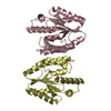
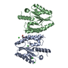
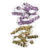
- PDB-2ccj: Crystal structure of S. aureus thymidylate kinase complexed with ... -
+ Open data

- Basic information
| Entry | Database: PDB / ID: 2ccj | ||||||
|---|---|---|---|---|---|---|---|
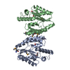
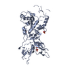
| Title | Crystal structure of S. aureus thymidylate kinase complexed with thymidine monophosphate | ||||||
 Components Components | THYMIDYLATE KINASE | ||||||
 Keywords Keywords | TRANSFERASE / KINASE / NUCLEOTIDE BIOSYNTHESIS / TMP-BINDING / ATP-BINDING / THYMIDYLATE KINASE | ||||||
| Function / homology |  Function and homology information Function and homology informationdTMP kinase / dUDP biosynthetic process / dTDP biosynthetic process / dTMP kinase activity / dTTP biosynthetic process / ATP binding / cytosol Similarity search - Function | ||||||
| Biological species |   STAPHYLOCOCCUS AUREUS (bacteria) STAPHYLOCOCCUS AUREUS (bacteria) | ||||||
| Method |  X-RAY DIFFRACTION / MOLECULAR REPLACEMENT / Resolution: 1.7 Å X-RAY DIFFRACTION / MOLECULAR REPLACEMENT / Resolution: 1.7 Å | ||||||
 Authors Authors | Kotaka, M. / Dhaliwal, B. / Ren, J. / Nichols, C.E. / Angell, R. / Lockyer, M. / Hawkins, A.R. / Stammers, D.K. | ||||||
 Citation Citation | Journal: Protein Sci. / Year: 2006 Title: Structures of S. Aureus Thymidylate Kinase Reveal an Atypical Active Site Configuration and an Intermediate Conformational State Upon Substrate Binding Authors: Kotaka, M. / Dhaliwal, B. / Ren, J. / Nichols, C.E. / Angell, R. / Lockyer, M. / Hawkins, A.R. / Stammers, D.K. | ||||||
| History |
|
- Structure visualization
| Structure viewer | Molecule: MolmilJmol/JSmol |
|---|
- Downloads & links
-Download
| PDBx/mmCIF format | 2ccj.cif.gz | 102.5 KB | Display | PDBx/mmCIF format |
|---|---|---|---|---|
| PDB format | pdb2ccj.ent.gz | 78.1 KB | Display | PDB format |
| PDBx/mmJSON format | 2ccj.json.gz | Tree view | PDBx/mmJSON format | |
| Others |  Other downloads Other downloads |
-Validation report
| Arichive directory | https://data.pdbj.org/pub/pdb/validation_reports/cc/2ccjftp://data.pdbj.org/pub/pdb/validation_reports/cc/2ccj | HTTPS FTP |
|---|
-Related structure data
| Related structure data | 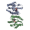 2ccgSC  2cckC S: Starting model for refinement C: citing same article ( |
|---|---|
| Similar structure data |


-Links
 PDBj
PDBj- Assembly
| Deposited unit | 
| ||||||||
|---|---|---|---|---|---|---|---|---|---|
| 1 |
| ||||||||
| Unit cell |
| ||||||||
| Noncrystallographic symmetry (NCS) | NCS oper: (Code: given Matrix: (-0.13987, 0.24966, 0.95818), Vector: |
-Components
| #1: Protein | Mass: 23454.586 Da / Num. of mol.: 2 Source method: isolated from a genetically manipulated source Source: (gene. exp.) STAPHYLOCOCCUS AUREUS (bacteria) / Plasmid: PET3A / Production host: #2: Chemical | ChemComp-CL /   Mass: 35.453 Da / Num. of mol.: 5 / Source method: obtained synthetically / Formula: Cl Mass: 35.453 Da / Num. of mol.: 5 / Source method: obtained synthetically / Formula: Cl#3: Chemical | 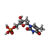  Mass: 322.208 Da / Num. of mol.: 3 / Source method: obtained synthetically / Formula: C10H15N2O8P Mass: 322.208 Da / Num. of mol.: 3 / Source method: obtained synthetically / Formula: C10H15N2O8P#4: Chemical |   Mass: 62.068 Da / Num. of mol.: 3 / Source method: obtained synthetically / Formula: C2H6O2 Mass: 62.068 Da / Num. of mol.: 3 / Source method: obtained synthetically / Formula: C2H6O2#5: Water | ChemComp-HOH / |  Mass: 18.015 Da / Num. of mol.: 413 / Source method: isolated from a natural source / Formula: H2O Mass: 18.015 Da / Num. of mol.: 413 / Source method: isolated from a natural source / Formula: H2O |
|---|
-Experimental details
-Experiment
| Experiment | Method: X-RAY DIFFRACTION / Number of used crystals: 1 |
|---|
- Sample preparation
| Crystal | Density Matthews: 2.17 Å3/Da / Density % sol: 42.98 % |
|---|---|
| Crystal grow | pH: 7.2 / Details: 1M LICL, 0.1M NA CACODYLATE PH 7.2, 18% PEG 6000 |
-Data collection
| Diffraction | Mean temperature: 105 K |
|---|---|
| Diffraction source | Source: ROTATING ANODE / Type: RIGAKU MICROMAX-007 / Wavelength: 1.5418 |
| Detector | Type: MARRESEARCH / Detector: IMAGE PLATE / Date: Feb 7, 2005 / Details: MIRRORS |
| Radiation | Protocol: SINGLE WAVELENGTH / Monochromatic (M) / Laue (L): M / Scattering type: x-ray |
| Radiation wavelength | Wavelength: 1.5418 Å / Relative weight: 1 |
| Reflection | Resolution: 1.7→30 Å / Num. obs: 43797 / % possible obs: 99.4 % / Observed criterion σ(I): 0 / Redundancy: 4.7 % / Biso Wilson estimate: 21.7 Å2 / Rmerge(I) obs: 0.04 / Net I/σ(I): 43.9 |
| Reflection shell | Resolution: 1.7→1.76 Å / Redundancy: 4.5 % / Rmerge(I) obs: 0.21 / Mean I/σ(I) obs: 7.1 / % possible all: 99.4 |
- Processing
| Software |
| ||||||||||||||||||||||||||||||||||||||||||||||||||||||||||||||||||||||||||||||||
|---|---|---|---|---|---|---|---|---|---|---|---|---|---|---|---|---|---|---|---|---|---|---|---|---|---|---|---|---|---|---|---|---|---|---|---|---|---|---|---|---|---|---|---|---|---|---|---|---|---|---|---|---|---|---|---|---|---|---|---|---|---|---|---|---|---|---|---|---|---|---|---|---|---|---|---|---|---|---|---|---|---|
| Refinement | Method to determine structure: MOLECULAR REPLACEMENT Starting model: PDB ENTRY 2CCG Resolution: 1.7→20.86 Å / Rfactor Rfree error: 0.005 / Data cutoff high absF: 745568.57 / Isotropic thermal model: RESTRAINED / Cross valid method: THROUGHOUT / σ(F): 0 / Stereochemistry target values: MAXIMUM LIKELIHOOD
| ||||||||||||||||||||||||||||||||||||||||||||||||||||||||||||||||||||||||||||||||
| Solvent computation | Solvent model: FLAT MODEL / Bsol: 56.4638 Å2 / ksol: 0.365152 e/Å3 | ||||||||||||||||||||||||||||||||||||||||||||||||||||||||||||||||||||||||||||||||
| Displacement parameters | Biso mean: 26.3 Å2
| ||||||||||||||||||||||||||||||||||||||||||||||||||||||||||||||||||||||||||||||||
| Refine analyze |
| ||||||||||||||||||||||||||||||||||||||||||||||||||||||||||||||||||||||||||||||||
| Refinement step | Cycle: LAST / Resolution: 1.7→20.86 Å
| ||||||||||||||||||||||||||||||||||||||||||||||||||||||||||||||||||||||||||||||||
| Refine LS restraints |
| ||||||||||||||||||||||||||||||||||||||||||||||||||||||||||||||||||||||||||||||||
| LS refinement shell | Resolution: 1.7→1.81 Å / Rfactor Rfree error: 0.013 / Total num. of bins used: 6
| ||||||||||||||||||||||||||||||||||||||||||||||||||||||||||||||||||||||||||||||||
| Xplor file |
|
