| 登録情報 | データベース: PDB / ID: 2pl0
|
|---|
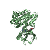
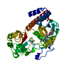
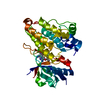
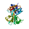
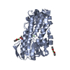
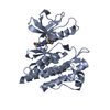
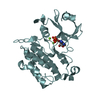
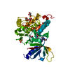
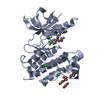
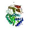
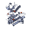
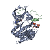
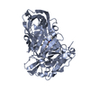
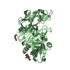
| タイトル | LCK bound to imatinib |
|---|
 要素 要素 | Proto-oncogene tyrosine-protein kinase LCK |
|---|
 キーワード キーワード | TRANSFERASE / kinase phosphorylation |
|---|
| 機能・相同性 |  機能・相同性情報 機能・相同性情報
regulation of lymphocyte activation / positive regulation of leukocyte cell-cell adhesion / CD27 signaling pathway / regulation of regulatory T cell differentiation / gamma-delta T cell differentiation / positive regulation of gamma-delta T cell differentiation / Fc-gamma receptor signaling pathway / FLT3 signaling through SRC family kinases / protein antigen binding / Nef Mediated CD4 Down-regulation ...regulation of lymphocyte activation / positive regulation of leukocyte cell-cell adhesion / CD27 signaling pathway / regulation of regulatory T cell differentiation / gamma-delta T cell differentiation / positive regulation of gamma-delta T cell differentiation / Fc-gamma receptor signaling pathway / FLT3 signaling through SRC family kinases / protein antigen binding / Nef Mediated CD4 Down-regulation / intracellular zinc ion homeostasis / CD4 receptor binding / positive regulation of heterotypic cell-cell adhesion / Nef and signal transduction / Co-stimulation by CD28 / Interleukin-2 signaling / CD28 dependent Vav1 pathway / peptidyl-tyrosine autophosphorylation / Regulation of KIT signaling / leukocyte migration / phospholipase activator activity / Co-inhibition by CTLA4 / CD8 receptor binding / Translocation of ZAP-70 to Immunological synapse / Phosphorylation of CD3 and TCR zeta chains / pericentriolar material / positive regulation of T cell receptor signaling pathway / protein serine/threonine phosphatase activity / PECAM1 interactions / hemopoiesis / Generation of second messenger molecules / RHOH GTPase cycle / T cell differentiation / immunological synapse / Co-inhibition by PD-1 / CD28 dependent PI3K/Akt signaling / T cell receptor binding / phosphatidylinositol 3-kinase binding / GPVI-mediated activation cascade / phospholipase binding / positive regulation of intrinsic apoptotic signaling pathway / T cell costimulation / phosphotyrosine residue binding / release of sequestered calcium ion into cytosol / SH2 domain binding / cell surface receptor protein tyrosine kinase signaling pathway / peptidyl-tyrosine phosphorylation / Signaling by phosphorylated juxtamembrane, extracellular and kinase domain KIT mutants / T cell activation / B cell receptor signaling pathway / non-membrane spanning protein tyrosine kinase activity / non-specific protein-tyrosine kinase / Signaling by SCF-KIT / positive regulation of T cell activation / platelet activation / Constitutive Signaling by Aberrant PI3K in Cancer / cell-cell junction / DAP12 signaling / Downstream TCR signaling / PIP3 activates AKT signaling / T cell receptor signaling pathway / ATPase binding / PI5P, PP2A and IER3 Regulate PI3K/AKT Signaling / protein tyrosine kinase activity / protein phosphatase binding / protein phosphorylation / intracellular signal transduction / membrane raft / response to xenobiotic stimulus / signaling receptor binding / positive regulation of gene expression / protein kinase binding / extracellular exosome / ATP binding / identical protein binding / plasma membrane / cytosol / cytoplasm類似検索 - 分子機能 Lck, SH3 domain / Tyrosine-protein kinase Lck, SH2 domain / : / SH3 domain / SH2 domain / Src homology 2 (SH2) domain profile. / Src homology 2 domains / SH2 domain / Src homology 3 domains / SH2 domain superfamily ...Lck, SH3 domain / Tyrosine-protein kinase Lck, SH2 domain / : / SH3 domain / SH2 domain / Src homology 2 (SH2) domain profile. / Src homology 2 domains / SH2 domain / Src homology 3 domains / SH2 domain superfamily / SH3-like domain superfamily / Src homology 3 (SH3) domain profile. / SH3 domain / Tyrosine-protein kinase, catalytic domain / Tyrosine kinase, catalytic domain / Tyrosine protein kinases specific active-site signature. / Tyrosine-protein kinase, active site / Serine-threonine/tyrosine-protein kinase, catalytic domain / Protein tyrosine and serine/threonine kinase / Phosphorylase Kinase; domain 1 / Phosphorylase Kinase; domain 1 / Transferase(Phosphotransferase) domain 1 / Transferase(Phosphotransferase); domain 1 / Protein kinase, ATP binding site / Protein kinases ATP-binding region signature. / Protein kinase domain profile. / Protein kinase domain / Protein kinase-like domain superfamily / 2-Layer Sandwich / Orthogonal Bundle / Mainly Alpha / Alpha Beta類似検索 - ドメイン・相同性 |
|---|
| 生物種 |  Homo sapiens (ヒト) Homo sapiens (ヒト) |
|---|
| 手法 |  X線回折 / シンクロトロン / 分子置換 / 解像度: 2.8 Å X線回折 / シンクロトロン / 分子置換 / 解像度: 2.8 Å |
|---|
 データ登録者 データ登録者 | Jacobs, M.D. |
|---|
 引用 引用 | ジャーナル: Proteins / 年: 2007
タイトル: Classifying protein kinase structures guides use of ligand-selectivity profiles to predict inactive conformations: Structure of lck/imatinib complex.
著者: Jacobs, M.D. / Caron, P.R. / Hare, B.J. |
|---|
| 履歴 | | 登録 | 2007年4月18日 | 登録サイト: RCSB / 処理サイト: RCSB |
|---|
| 改定 1.0 | 2007年10月9日 | Provider: repository / タイプ: Initial release |
|---|
| 改定 1.1 | 2011年7月13日 | Group: Version format compliance |
|---|
| 改定 1.2 | 2024年2月21日 | Group: Data collection / Database references ...Data collection / Database references / Derived calculations / Structure summary
カテゴリ: chem_comp / chem_comp_atom ...chem_comp / chem_comp_atom / chem_comp_bond / database_2 / struct_ref_seq_dif / struct_site
Item: _chem_comp.pdbx_synonyms / _database_2.pdbx_DOI ..._chem_comp.pdbx_synonyms / _database_2.pdbx_DOI / _database_2.pdbx_database_accession / _struct_ref_seq_dif.details / _struct_site.pdbx_auth_asym_id / _struct_site.pdbx_auth_comp_id / _struct_site.pdbx_auth_seq_id |
|---|
|
|---|
 ムービー
ムービー コントローラー


 データを開く
 基本情報
基本情報 構造の表示
構造の表示 ダウンロードとリンク
 その他のダウンロード
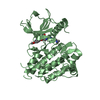


 PDBj
PDBj


 集合体
集合体

 Spodoptera frugiperda (ツマジロクサヨトウ)
 分子量: 493.603 Da / 分子数: 1 / 由来タイプ: 合成 / 式: C29H31N7O / コメント: 薬剤*YM
 分子量: 18.015 Da / 分子数: 93 / 由来タイプ: 天然 / 式: H2O
 試料調製
試料調製 / ビームライン: 5.0.2 / 波長: 1 Å
 解析
解析