[English] 日本語
Yorodumi
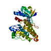
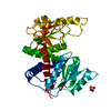
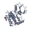
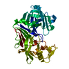
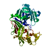
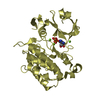
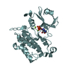
- PDB-3dls: Crystal structure of human PAS kinase bound to ADP -

+
Open data


ID or keywords:

Loading...

-
Basic information

Entry
Database: PDB / ID: 3dls
TitleCrystal structure of human PAS kinase bound to ADP
ComponentsPAS domain-containing serine/threonine-protein kinase
KeywordsTRANSFERASE / PAS KINASE / PASK / PROTEIN KINASE / DRUG DISCOVERY / ATP-binding / Kinase / Nucleotide-binding / Phosphoprotein / Serine/threonine-protein kinase / Structural Genomics / PSI-2 / Protein Structure Initiative / New York SGX Research Center for Structural Genomics / NYSGXRC
Function / homology
Function and homology information


regulation of glucagon secretion / negative regulation of glycogen biosynthetic process / regulation of respiratory gaseous exchange / energy homeostasis / phosphatidylinositol binding / positive regulation of translation / protein autophosphorylation / non-specific serine/threonine protein kinase / intracellular signal transduction / protein phosphorylation ...regulation of glucagon secretion / negative regulation of glycogen biosynthetic process / regulation of respiratory gaseous exchange / energy homeostasis / phosphatidylinositol binding / positive regulation of translation / protein autophosphorylation / non-specific serine/threonine protein kinase / intracellular signal transduction / protein phosphorylation / protein serine kinase activity / protein serine/threonine kinase activity / ATP binding / nucleus / cytoplasm / cytosol
Similarity search - Function
PAS domain / PAS domain / PAS repeat profile. / PAS domain / PAS domain superfamily / Transferase(Phosphotransferase) domain 1 / Transferase(Phosphotransferase); domain 1 / Phosphorylase Kinase; domain 1 / Phosphorylase Kinase; domain 1 / Serine/threonine-protein kinase, active site ...PAS domain / PAS domain / PAS repeat profile. / PAS domain / PAS domain superfamily / Transferase(Phosphotransferase) domain 1 / Transferase(Phosphotransferase); domain 1 / Phosphorylase Kinase; domain 1 / Phosphorylase Kinase; domain 1 / Serine/threonine-protein kinase, active site / Serine/Threonine protein kinases active-site signature. / Protein kinase domain / Serine/Threonine protein kinases, catalytic domain / Protein kinase, ATP binding site / Protein kinases ATP-binding region signature. / Protein kinase domain profile. / Protein kinase domain / Protein kinase-like domain superfamily / 2-Layer Sandwich / Orthogonal Bundle / Mainly Alpha / Alpha Beta
Similarity search - Domain/homology
ADENOSINE-5'-DIPHOSPHATE / PAS domain-containing serine/threonine-protein kinase
Similarity search - Component
Biological speciesHomo sapiens (human)
MethodX-RAY DIFFRACTION / SYNCHROTRON / Resolution: 2.3 Å
AuthorsAntonysamy, S. / Bonanno, J.B. / Romero, R. / Russell, M. / Iizuka, M. / Gheyi, T. / Wasserman, S.R. / Rutter, J. / Sauder, J.M. / Burley, S.K. / New York SGX Research Center for Structural Genomics (NYSGXRC)
CitationJournal: J.Biol.Chem. / Year: 2010
Title: Structural bases of PAS domain-regulated kinase (PASK) activation in the absence of activation loop phosphorylation.
Authors: Kikani, C.K. / Antonysamy, S.A. / Bonanno, J.B. / Romero, R. / Zhang, F.F. / Russell, M. / Gheyi, T. / Iizuka, M. / Emtage, S. / Sauder, J.M. / Turk, B.E. / Burley, S.K. / Rutter, J.
History
DepositionJun 29, 2008Deposition site: RCSB / Processing site: RCSB
Revision 1.0Aug 26, 2008Provider: repository / Type: Initial release
Revision 1.1Jul 13, 2011Group: Derived calculations / Version format compliance
Revision 1.2Aug 19, 2015Group: Database references
Revision 1.3Nov 14, 2018Group: Data collection / Structure summary / Category: audit_author / Item: _audit_author.identifier_ORCID
Revision 1.4Feb 10, 2021Group: Database references / Derived calculations / Structure summary
Category: audit_author / citation_author ...audit_author / citation_author / pdbx_struct_conn_angle / struct_conn / struct_site
Item: _audit_author.identifier_ORCID / _citation_author.identifier_ORCID ..._audit_author.identifier_ORCID / _citation_author.identifier_ORCID / _pdbx_struct_conn_angle.ptnr1_auth_asym_id / _pdbx_struct_conn_angle.ptnr1_auth_comp_id / _pdbx_struct_conn_angle.ptnr1_auth_seq_id / _pdbx_struct_conn_angle.ptnr1_label_asym_id / _pdbx_struct_conn_angle.ptnr1_label_atom_id / _pdbx_struct_conn_angle.ptnr1_label_comp_id / _pdbx_struct_conn_angle.ptnr1_label_seq_id / _pdbx_struct_conn_angle.ptnr2_auth_asym_id / _pdbx_struct_conn_angle.ptnr2_auth_seq_id / _pdbx_struct_conn_angle.ptnr2_label_asym_id / _pdbx_struct_conn_angle.ptnr3_auth_asym_id / _pdbx_struct_conn_angle.ptnr3_auth_comp_id / _pdbx_struct_conn_angle.ptnr3_auth_seq_id / _pdbx_struct_conn_angle.ptnr3_label_asym_id / _pdbx_struct_conn_angle.ptnr3_label_atom_id / _pdbx_struct_conn_angle.ptnr3_label_comp_id / _pdbx_struct_conn_angle.ptnr3_label_seq_id / _pdbx_struct_conn_angle.value / _struct_conn.pdbx_dist_value / _struct_conn.ptnr1_auth_asym_id / _struct_conn.ptnr1_auth_comp_id / _struct_conn.ptnr1_auth_seq_id / _struct_conn.ptnr1_label_asym_id / _struct_conn.ptnr1_label_atom_id / _struct_conn.ptnr1_label_comp_id / _struct_conn.ptnr1_label_seq_id / _struct_conn.ptnr2_auth_asym_id / _struct_conn.ptnr2_auth_comp_id / _struct_conn.ptnr2_auth_seq_id / _struct_conn.ptnr2_label_asym_id / _struct_conn.ptnr2_label_atom_id / _struct_conn.ptnr2_label_comp_id / _struct_conn.ptnr2_label_seq_id / _struct_site.pdbx_auth_asym_id / _struct_site.pdbx_auth_comp_id / _struct_site.pdbx_auth_seq_id
Revision 1.5Feb 21, 2024Group: Data collection / Database references / Category: chem_comp_atom / chem_comp_bond / database_2
Item: _database_2.pdbx_DOI / _database_2.pdbx_database_accession

-
Structure visualization

Structure viewerMolecule:
MolmilJmol/JSmol

Downloads & links

-
Assembly

Deposited unit
A: PAS domain-containing serine/threonine-protein kinase
B: PAS domain-containing serine/threonine-protein kinase
C: PAS domain-containing serine/threonine-protein kinase
D: PAS domain-containing serine/threonine-protein kinase
E: PAS domain-containing serine/threonine-protein kinase
F: PAS domain-containing serine/threonine-protein kinase
hetero molecules


Theoretical massNumber of molelcules
Total (without water)230,27731
Polymers227,2526
Non-polymers3,02525
Water5,044280
1
A: PAS domain-containing serine/threonine-protein kinase
hetero molecules


Theoretical massNumber of molelcules
Total (without water)38,4006
Polymers37,8751
Non-polymers5245
Water181
TypeNameSymmetry operationNumber
identity operation1_555x,y,z1
MethodPQS
2
B: PAS domain-containing serine/threonine-protein kinase
hetero molecules


Theoretical massNumber of molelcules
Total (without water)38,4006
Polymers37,8751
Non-polymers5245
Water181
TypeNameSymmetry operationNumber
identity operation1_555x,y,z1
MethodPQS
3
C: PAS domain-containing serine/threonine-protein kinase
hetero molecules


Theoretical massNumber of molelcules
Total (without water)38,4006
Polymers37,8751
Non-polymers5245
Water181
TypeNameSymmetry operationNumber
identity operation1_555x,y,z1
MethodPQS
4
D: PAS domain-containing serine/threonine-protein kinase
hetero molecules


Theoretical massNumber of molelcules
Total (without water)38,3755
Polymers37,8751
Non-polymers5004
Water181
TypeNameSymmetry operationNumber
identity operation1_555x,y,z1
MethodPQS
5
E: PAS domain-containing serine/threonine-protein kinase
hetero molecules


Theoretical massNumber of molelcules
Total (without water)38,3514
Polymers37,8751
Non-polymers4763
Water181
TypeNameSymmetry operationNumber
identity operation1_555x,y,z1
MethodPQS
6
F: PAS domain-containing serine/threonine-protein kinase
hetero molecules


Theoretical massNumber of molelcules
Total (without water)38,3514
Polymers37,8751
Non-polymers4763
Water181
TypeNameSymmetry operationNumber
identity operation1_555x,y,z1
MethodPQS
7
A: PAS domain-containing serine/threonine-protein kinase
B: PAS domain-containing serine/threonine-protein kinase
C: PAS domain-containing serine/threonine-protein kinase
hetero molecules


Theoretical massNumber of molelcules
Total (without water)115,19918
Polymers113,6263
Non-polymers1,57315
Water543
TypeNameSymmetry operationNumber
identity operation1_555x,y,z1
Buried area7200 Å2
ΔGint-162 kcal/mol
Surface area38530 Å2
MethodPISA
8
D: PAS domain-containing serine/threonine-protein kinase
hetero molecules

E: PAS domain-containing serine/threonine-protein kinase
hetero molecules

F: PAS domain-containing serine/threonine-protein kinase
hetero molecules


Theoretical massNumber of molelcules
Total (without water)115,07813
Polymers113,6263
Non-polymers1,45210
Water543
TypeNameSymmetry operationNumber
identity operation1_555x,y,z1
crystal symmetry operation1_565x,y+1,z1
crystal symmetry operation1_465x-1,y+1,z1
Buried area6620 Å2
ΔGint-123 kcal/mol
Surface area37970 Å2
MethodPISA
Unit cell
Length a, b, c (Å)85.778, 85.840, 94.154
Angle α, β, γ (deg.)77.280, 77.500, 60.090
Int Tables number1
Space group name H-MP1

-
Components

#1: Protein
PAS domain-containing serine/threonine-protein kinase / PAS-kinase / PASKIN / hPASK


Mass: 37875.270 Da / Num. of mol.: 6 / Fragment: PROTEIN KINASE DOMAIN
Source method: isolated from a genetically manipulated source
Source: (gene. exp.) Homo sapiens (human) / Gene: PASK, KIAA0135 / Production host: 
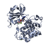
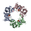
BACULOVIRUS / Strain (production host): SF9
References: UniProt: Q96RG2, non-specific serine/threonine protein kinase
#2: Chemical
ChemComp-MG / MAGNESIUM ION


Mass: 24.305 Da / Num. of mol.: 19 / Source method: obtained synthetically / Formula: Mg
#3: Chemical
ChemComp-ADP / ADENOSINE-5'-DIPHOSPHATE


Mass: 427.201 Da / Num. of mol.: 6 / Source method: obtained synthetically / Formula: C10H15N5O10P2 / Comment: ADP, energy-carrying molecule*YM
#4: Water ChemComp-HOH / water


Mass: 18.015 Da / Num. of mol.: 280 / Source method: isolated from a natural source / Formula: H2O

-
Experimental details

-
Experiment

ExperimentMethod: X-RAY DIFFRACTION / Number of used crystals: 1

-
Sample preparation

CrystalDensity Matthews: 2.56 Å3/Da / Density % sol: 51.93 %
Crystal growTemperature: 293 K / Method: vapor diffusion / pH: 7.5
Details: 10% GLYCEROL, 18% PEG 3350, 150 mM CALCIUM ACETATE, pH 7.5, VAPOR DIFFUSION, temperature 293K

-
Data collection

DiffractionMean temperature: 100 K
Diffraction sourceSource: SYNCHROTRON / Site: APS / Beamline: 31-ID / Wavelength: 0.97929 Å
DetectorType: MAR CCD 165 mm / Detector: CCD / Date: Apr 22, 2008
RadiationMonochromator: DIAMOND / Protocol: SINGLE WAVELENGTH / Monochromatic (M) / Laue (L): M / Scattering type: x-ray
Radiation wavelengthWavelength: 0.97929 Å / Relative weight: 1
ReflectionHighest resolution: 2.3 Å / Num. obs: 96809

-
Processing

Software
NameVersionClassificationNB
REFMACrefinement
PDB_EXTRACT3.006data extraction
RefinementHighest resolution: 2.3 Å / Occupancy max: 1 / Occupancy min: 1
RfactorNum. reflection
Rfree0.297 -
Rwork0.241 -
all0.244 96809
obs0.244 96809
Displacement parametersBiso max: 81.99 Å2 / Biso mean: 37.365 Å2 / Biso min: 2.72 Å2
Refinement stepCycle: LAST / Highest resolution: 2.3 Å
ProteinNucleic acidLigandSolventTotal
Num. atoms13263 0 181 280 13724

+
About Yorodumi

-
News

-
Feb 9, 2022. New format data for meta-information of EMDB entries

New format data for meta-information of EMDB entries

  • Version 3 of the EMDB header file is now the official format.
  • The previous official version 1.9 will be removed from the archive.

Related info.:EMDB header

External links:wwPDB to switch to version 3 of the EMDB data model

-
Aug 12, 2020. Covid-19 info

Covid-19 info

URL: https://pdbjlvh1.pdbj.org/emnavi/covid19.php

New page: Covid-19 featured information page in EM Navigator.

Related info.:Covid-19 info / Mar 5, 2020. Novel coronavirus structure data

+
Mar 5, 2020. Novel coronavirus structure data

Novel coronavirus structure data

Related info.:Yorodumi Speices / Aug 12, 2020. Covid-19 info

External links:COVID-19 featured content - PDBj / Molecule of the Month (242):Coronavirus Proteases

+
Jan 31, 2019. EMDB accession codes are about to change! (news from PDBe EMDB page)

EMDB accession codes are about to change! (news from PDBe EMDB page)

  • The allocation of 4 digits for EMDB accession codes will soon come to an end. Whilst these codes will remain in use, new EMDB accession codes will include an additional digit and will expand incrementally as the available range of codes is exhausted. The current 4-digit format prefixed with “EMD-” (i.e. EMD-XXXX) will advance to a 5-digit format (i.e. EMD-XXXXX), and so on. It is currently estimated that the 4-digit codes will be depleted around Spring 2019, at which point the 5-digit format will come into force.
  • The EM Navigator/Yorodumi systems omit the EMD- prefix.

Related info.:Q: What is EMD? / ID/Accession-code notation in Yorodumi/EM Navigator

External links:EMDB Accession Codes are Changing Soon! / Contact to PDBj

+
Jul 12, 2017. Major update of PDB

Major update of PDB

  • wwPDB released updated PDB data conforming to the new PDBx/mmCIF dictionary.
  • This is a major update changing the version number from 4 to 5, and with Remediation, in which all the entries are updated.
  • In this update, many items about electron microscopy experimental information are reorganized (e.g. em_software).
  • Now, EM Navigator and Yorodumi are based on the updated data.

External links:wwPDB Remediation / Enriched Model Files Conforming to OneDep Data Standards Now Available in the PDB FTP Archive

-
Yorodumi

Thousand views of thousand structures

  • Yorodumi is a browser for structure data from EMDB, PDB, SASBDB, etc.
  • This page is also the successor to EM Navigator detail page, and also detail information page/front-end page for Omokage search.
  • The word "yorodu" (or yorozu) is an old Japanese word meaning "ten thousand". "mi" (miru) is to see.

Related info.:EMDB / PDB / SASBDB / Comparison of 3 databanks / Yorodumi Search / Aug 31, 2016. New EM Navigator & Yorodumi / Yorodumi Papers / Jmol/JSmol / Function and homology information / Changes in new EM Navigator and Yorodumi

Read more