 Movie
Movie Controller

[English] 日本語
 Yorodumi
- PDB-1pvv: Refined Structure of Pyrococcus furiosus Ornithine Carbamoyltrans... -
+ Open data

- Basic information
| Entry | Database: PDB / ID: 1pvv | ||||||
|---|---|---|---|---|---|---|---|
| Title | Refined Structure of Pyrococcus furiosus Ornithine Carbamoyltransferase at 1.87 A | ||||||
 Components Components | Ornithine carbamoyltransferase | ||||||
 Keywords Keywords | TRANSFERASE / dodecamer | ||||||
| Function / homology |  Function and homology information Function and homology informationornithine carbamoyltransferase / ornithine carbamoyltransferase activity / L-citrulline biosynthetic process / : / amino acid binding / cytoplasm Similarity search - Function | ||||||
| Biological species |   Pyrococcus furiosus (archaea) Pyrococcus furiosus (archaea) | ||||||
| Method |  X-RAY DIFFRACTION / SYNCHROTRON / AB INITIO / Resolution: 1.87 Å X-RAY DIFFRACTION / SYNCHROTRON / AB INITIO / Resolution: 1.87 Å | ||||||
 Authors Authors | Massant, J. / Wouters, J. / Glansdorff, N. | ||||||
 Citation Citation | Journal: Acta Crystallogr.,Sect.D / Year: 2003 Title: Refined structure of Pyrococcus furiosus ornithine carbamoyltransferase at 1.87 A. Authors: Massant, J. / Wouters, J. / Glansdorff, N. | ||||||
| History |
|
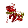
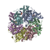
- Structure visualization
| Structure viewer | Molecule: MolmilJmol/JSmol |
|---|
- Downloads & links
-Download
| PDBx/mmCIF format | 1pvv.cif.gz | 79.6 KB | Display | PDBx/mmCIF format |
|---|---|---|---|---|
| PDB format | pdb1pvv.ent.gz | 58.7 KB | Display | PDB format |
| PDBx/mmJSON format | 1pvv.json.gz | Tree view | PDBx/mmJSON format | |
| Others |  Other downloads Other downloads |
-Validation report
| Arichive directory | https://data.pdbj.org/pub/pdb/validation_reports/pv/1pvvftp://data.pdbj.org/pub/pdb/validation_reports/pv/1pvv | HTTPS FTP |
|---|
-Related structure data
| Related structure data |  1a1sS S: Starting model for refinement |
|---|---|
| Similar structure data |


-Links
 PDBj
PDBj
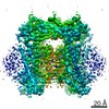
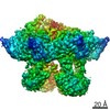
- Assembly
| Deposited unit | 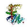
| |||||||||||||||||||||||||||
|---|---|---|---|---|---|---|---|---|---|---|---|---|---|---|---|---|---|---|---|---|---|---|---|---|---|---|---|---|
| 1 | x 12
| |||||||||||||||||||||||||||
| Unit cell |
| |||||||||||||||||||||||||||
| Components on special symmetry positions |
| |||||||||||||||||||||||||||
| Details | The biological assembly is a dodecamer generated from the monomer in the asymmetric unit |
-Components
| #1: Protein | Mass: 35229.387 Da / Num. of mol.: 1 Source method: isolated from a genetically manipulated source Source: (gene. exp.) Pyrococcus furiosus (archaea) / Gene: ARGF OR PF0594 / Production host:  | ||
|---|---|---|---|
| #2: Chemical |   Mass: 96.063 Da / Num. of mol.: 2 / Source method: obtained synthetically / Formula: SO4 Mass: 96.063 Da / Num. of mol.: 2 / Source method: obtained synthetically / Formula: SO4#3: Water | ChemComp-HOH / |  Mass: 18.015 Da / Num. of mol.: 191 / Source method: isolated from a natural source / Formula: H2O Mass: 18.015 Da / Num. of mol.: 191 / Source method: isolated from a natural source / Formula: H2O |
-Experimental details
-Experiment
| Experiment | Method: X-RAY DIFFRACTION / Number of used crystals: 1 |
|---|
- Sample preparation
| Crystal | Density Matthews: 3.23 Å3/Da / Density % sol: 67.02 % | ||||||||||||||||||||||||||||||||||||
|---|---|---|---|---|---|---|---|---|---|---|---|---|---|---|---|---|---|---|---|---|---|---|---|---|---|---|---|---|---|---|---|---|---|---|---|---|---|
| Crystal grow | Temperature: 293 K / Method: vapor diffusion, hanging drop / pH: 4.6 Details: sodium acetate, ammonium sulfate, glycerol, pH 4.6, VAPOR DIFFUSION, HANGING DROP, temperature 293K | ||||||||||||||||||||||||||||||||||||
| Crystal grow | *PLUS Method: vapor diffusion, hanging drop | ||||||||||||||||||||||||||||||||||||
| Components of the solutions | *PLUS
|
-Data collection
| Diffraction | Mean temperature: 100 K |
|---|---|
| Diffraction source | Source: SYNCHROTRON / Site: ESRF  / Beamline: BM30A / Wavelength: 0.989 Å / Beamline: BM30A / Wavelength: 0.989 Å |
| Detector | Type: MARRESEARCH / Detector: CCD / Date: Sep 1, 2001 |
| Radiation | Protocol: SINGLE WAVELENGTH / Monochromatic (M) / Laue (L): M / Scattering type: x-ray |
| Radiation wavelength | Wavelength: 0.989 Å / Relative weight: 1 |
| Reflection | Resolution: 1.87→99 Å / Num. all: 42928 / Num. obs: 42928 / % possible obs: 98.5 % / Rmerge(I) obs: 0.033 |
| Reflection shell | Resolution: 1.87→1.91 Å / Rmerge(I) obs: 0.187 / % possible all: 86.5 |
| Reflection | *PLUS Num. measured all: 304009 |
| Reflection shell | *PLUS % possible obs: 86.5 % |
- Processing
| Software |
| |||||||||||||||||||||||||||||||||
|---|---|---|---|---|---|---|---|---|---|---|---|---|---|---|---|---|---|---|---|---|---|---|---|---|---|---|---|---|---|---|---|---|---|---|
| Refinement | Method to determine structure: AB INITIO Starting model: PDB ENTRY 1A1S Resolution: 1.87→99 Å / Num. parameters: 10575 / Num. restraintsaints: 10110 / Cross valid method: FREE R / σ(F): 0 / Stereochemistry target values: ENGH AND HUBER
| |||||||||||||||||||||||||||||||||
| Refine analyze | Num. disordered residues: 0 / Occupancy sum hydrogen: 0 / Occupancy sum non hydrogen: 2648.48 | |||||||||||||||||||||||||||||||||
| Refinement step | Cycle: LAST / Resolution: 1.87→99 Å
| |||||||||||||||||||||||||||||||||
| Refine LS restraints |
| |||||||||||||||||||||||||||||||||
| LS refinement shell |
|
