 Movie
Movie Controller

[English] 日本語
 Yorodumi
- PDB-1vlv: Crystal structure of Ornithine carbamoyltransferase (TM1097) from... -
+ Open data

- Basic information
| Entry | Database: PDB / ID: 1vlv | ||||||
|---|---|---|---|---|---|---|---|
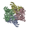
| Title | Crystal structure of Ornithine carbamoyltransferase (TM1097) from Thermotoga maritima at 2.25 A resolution | ||||||
 Components Components | Ornithine carbamoyltransferase | ||||||
 Keywords Keywords | TRANSFERASE / TM1097 / ORNITHINE CARBAMOYLTRANSFERASE / STRUCTURAL GENOMICS / JCSG / PROTEIN STRUCTURE INITIATIVE / PSI / Joint Center for Structural Genomics | ||||||
| Function / homology |  Function and homology information Function and homology informationornithine carbamoyltransferase / ornithine carbamoyltransferase activity / L-citrulline biosynthetic process / : / amino acid binding / cytoplasm Similarity search - Function | ||||||
| Biological species |   Thermotoga maritima (bacteria) Thermotoga maritima (bacteria) | ||||||
| Method |  X-RAY DIFFRACTION / SYNCHROTRON / MOLECULAR REPLACEMENT / Resolution: 2.25 Å X-RAY DIFFRACTION / SYNCHROTRON / MOLECULAR REPLACEMENT / Resolution: 2.25 Å | ||||||
 Authors Authors | Joint Center for Structural Genomics (JCSG) | ||||||
 Citation Citation | Journal: To be published Title: Crystal structure of Ornithine carbamoyltransferase (TM1097) from Thermotoga maritima at 2.25 A resolution Authors: Joint Center for Structural Genomics (JCSG) | ||||||
| History |
|
- Structure visualization
| Structure viewer | Molecule: MolmilJmol/JSmol |
|---|
- Downloads & links
-Download
| PDBx/mmCIF format | 1vlv.cif.gz | 77.3 KB | Display | PDBx/mmCIF format |
|---|---|---|---|---|
| PDB format | pdb1vlv.ent.gz | 58.6 KB | Display | PDB format |
| PDBx/mmJSON format | 1vlv.json.gz | Tree view | PDBx/mmJSON format | |
| Others |  Other downloads Other downloads |
-Validation report
| Arichive directory | https://data.pdbj.org/pub/pdb/validation_reports/vl/1vlvftp://data.pdbj.org/pub/pdb/validation_reports/vl/1vlv | HTTPS FTP |
|---|
-Related structure data
| Related structure data |  1a1sS S: Starting model for refinement |
|---|---|
| Similar structure data | |
| Other databases |

-Links
 PDBj
PDBj
- Assembly
| Deposited unit | 
| ||||||||||||||||||
|---|---|---|---|---|---|---|---|---|---|---|---|---|---|---|---|---|---|---|---|
| 1 | x 12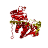
| ||||||||||||||||||
| Unit cell |
| ||||||||||||||||||
| Components on special symmetry positions |
|
-Components
| #1: Protein | Mass: 36870.402 Da / Num. of mol.: 1 Source method: isolated from a genetically manipulated source Source: (gene. exp.) Thermotoga maritima (bacteria) / Gene: argF / Production host: | ||
|---|---|---|---|
| #2: Chemical |   Mass: 94.971 Da / Num. of mol.: 3 / Source method: obtained synthetically / Formula: PO4 Mass: 94.971 Da / Num. of mol.: 3 / Source method: obtained synthetically / Formula: PO4#3: Water | ChemComp-HOH / |  Mass: 18.015 Da / Num. of mol.: 94 / Source method: isolated from a natural source / Formula: H2O Mass: 18.015 Da / Num. of mol.: 94 / Source method: isolated from a natural source / Formula: H2O |
-Experimental details
-Experiment
| Experiment | Method: X-RAY DIFFRACTION / Number of used crystals: 1 |
|---|
- Sample preparation
| Crystal | Density Matthews: 2.75 Å3/Da / Density % sol: 54.99 % |
|---|---|
| Crystal grow | Temperature: 293 K / Method: vapor diffusion, sitting drop, nanodrop / pH: 6.1 Details: 0.2% MPD, 0.2M NH4OAc, 0.1M Na,K-Phosphate pH 6.1, VAPOR DIFFUSION,SITTING DROP,NANODROP, temperature 293K |
-Data collection
| Diffraction | Mean temperature: 100 K |
|---|---|
| Diffraction source | Source: SYNCHROTRON / Site: ALS  / Beamline: 8.2.1 / Wavelength: 1 / Beamline: 8.2.1 / Wavelength: 1 |
| Detector | Type: ADSC / Detector: CCD / Date: Sep 20, 2003 |
| Radiation | Monochromator: Double Crystal Si(111) / Protocol: SINGLE WAVELENGTH / Monochromatic (M) / Laue (L): M / Scattering type: x-ray |
| Radiation wavelength | Wavelength: 1 Å / Relative weight: 1 |
| Reflection | Resolution: 2.25→63.32 Å / Num. obs: 19349 / % possible obs: 99.8 % / Redundancy: 6.9 % / Biso Wilson estimate: 58.84 Å2 / Rsym value: 0.071 / Net I/σ(I): 15.7 |
| Reflection shell | Resolution: 2.25→2.31 Å / Redundancy: 4.2 % / Mean I/σ(I) obs: 5014 / Num. unique all: 1366 / Rsym value: 0.734 / % possible all: 98.1 |
- Processing
| Software |
| ||||||||||||||||||||||||||||||||||||||||||||||||||||||||||||||||||||||||||||||||||||||||||||||||||||||||||||||||||||||||||||||||||||||||||||||||||||||
|---|---|---|---|---|---|---|---|---|---|---|---|---|---|---|---|---|---|---|---|---|---|---|---|---|---|---|---|---|---|---|---|---|---|---|---|---|---|---|---|---|---|---|---|---|---|---|---|---|---|---|---|---|---|---|---|---|---|---|---|---|---|---|---|---|---|---|---|---|---|---|---|---|---|---|---|---|---|---|---|---|---|---|---|---|---|---|---|---|---|---|---|---|---|---|---|---|---|---|---|---|---|---|---|---|---|---|---|---|---|---|---|---|---|---|---|---|---|---|---|---|---|---|---|---|---|---|---|---|---|---|---|---|---|---|---|---|---|---|---|---|---|---|---|---|---|---|---|---|---|---|---|
| Refinement | Method to determine structure: MOLECULAR REPLACEMENT Starting model: 1a1s Resolution: 2.25→63.32 Å / Cor.coef. Fo:Fc: 0.955 / Cor.coef. Fo:Fc free: 0.955 / SU B: 12.355 / SU ML: 0.147 / TLS residual ADP flag: LIKELY RESIDUAL / Cross valid method: THROUGHOUT / ESU R: 0.242 / ESU R Free: 0.184 / Stereochemistry target values: MAXIMUM LIKELIHOOD Details: 1), HYDROGENS HAVE BEEN ADDED IN THE RIDING POSITIONS. 2), THE DENSITY BETWEEN 235-244 IS WEAK AND IT IS NOT POSSIBLE TO BUILD THE MISSING RESIDUES INTO IT. 3), TWO PO4 ANIONS ARE LOCATED AT ...Details: 1), HYDROGENS HAVE BEEN ADDED IN THE RIDING POSITIONS. 2), THE DENSITY BETWEEN 235-244 IS WEAK AND IT IS NOT POSSIBLE TO BUILD THE MISSING RESIDUES INTO IT. 3), TWO PO4 ANIONS ARE LOCATED AT THE THREE-FOLD CRYSTALLOGRAPHIC SYMMETRY AXIS AT THE INTERFACE BETWEEN THREE MONOMERS. THE P AND O ATOMS ON THESE RESIDUES WERE MODELED WITH OCCUPANCIES OF 0.33.
| ||||||||||||||||||||||||||||||||||||||||||||||||||||||||||||||||||||||||||||||||||||||||||||||||||||||||||||||||||||||||||||||||||||||||||||||||||||||
| Solvent computation | Ion probe radii: 0.8 Å / Shrinkage radii: 0.8 Å / VDW probe radii: 1.2 Å / Solvent model: BABINET MODEL WITH MASK | ||||||||||||||||||||||||||||||||||||||||||||||||||||||||||||||||||||||||||||||||||||||||||||||||||||||||||||||||||||||||||||||||||||||||||||||||||||||
| Displacement parameters | Biso mean: 47.765 Å2 | ||||||||||||||||||||||||||||||||||||||||||||||||||||||||||||||||||||||||||||||||||||||||||||||||||||||||||||||||||||||||||||||||||||||||||||||||||||||
| Refinement step | Cycle: LAST / Resolution: 2.25→63.32 Å
| ||||||||||||||||||||||||||||||||||||||||||||||||||||||||||||||||||||||||||||||||||||||||||||||||||||||||||||||||||||||||||||||||||||||||||||||||||||||
| Refine LS restraints |
| ||||||||||||||||||||||||||||||||||||||||||||||||||||||||||||||||||||||||||||||||||||||||||||||||||||||||||||||||||||||||||||||||||||||||||||||||||||||
| LS refinement shell | Resolution: 2.25→2.313 Å / Total num. of bins used: 20
| ||||||||||||||||||||||||||||||||||||||||||||||||||||||||||||||||||||||||||||||||||||||||||||||||||||||||||||||||||||||||||||||||||||||||||||||||||||||
| Refinement TLS params. | Method: refined / Refine-ID: X-RAY DIFFRACTION
| ||||||||||||||||||||||||||||||||||||||||||||||||||||||||||||||||||||||||||||||||||||||||||||||||||||||||||||||||||||||||||||||||||||||||||||||||||||||
| Refinement TLS group | Refine-ID: X-RAY DIFFRACTION / Selection: ALL / Auth asym-ID: A / Label asym-ID: A
|
