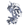
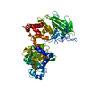
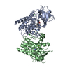
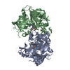
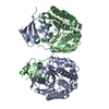
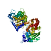
- PDB-2pi7: Structure of the catalytic domain of the chick retinal neurite in... -
+
データを開く
IDまたはキーワード:
読み込み中...
-
基本情報
登録情報
データベース: PDB / ID: 2pi7
タイトル
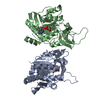
Structure of the catalytic domain of the chick retinal neurite inhibitor-Receptor Protein Tyrosine Phosphatase CRYP-2/cPTPRO
要素
Protein tyrosine phosphatase CRYP-2
キーワード
HYDROLASE / Protein Tyrosine Phosphatase
機能・相同性
機能・相同性情報
slit diaphragm assembly / regulation of glomerular filtration / negative regulation of retinal ganglion cell axon guidance / podocyte differentiation / Wnt-protein binding / glomerulus development / motor neuron axon guidance / lamellipodium assembly / negative regulation of glomerular filtration / peptidyl-tyrosine dephosphorylation ...slit diaphragm assembly / regulation of glomerular filtration / negative regulation of retinal ganglion cell axon guidance / podocyte differentiation / Wnt-protein binding / glomerulus development / motor neuron axon guidance / lamellipodium assembly / negative regulation of glomerular filtration / peptidyl-tyrosine dephosphorylation / monocyte chemotaxis / negative regulation of cell-substrate adhesion / lateral plasma membrane / protein-tyrosine-phosphatase / protein tyrosine phosphatase activity / axon guidance / negative regulation of canonical Wnt signaling pathway / cell morphogenesis / lamellipodium / negative regulation of neuron projection development / growth cone / dendritic spine / apical plasma membrane / neuron projection / axon / protein homodimerization activity / plasma membrane 類似検索 - 分子機能
Receptor-type tyrosine-protein phosphatase O / : / R-PTP-O-like, fibronectin type III domain / Protein tyrosine phosphatase superfamily / Protein-Tyrosine Phosphatase; Chain A / Protein tyrosine phosphatase, catalytic domain / PTP type protein phosphatase domain profile. / Protein-tyrosine phosphatase / Tyrosine-specific protein phosphatase, PTPase domain / Protein-tyrosine phosphatase, catalytic ...Receptor-type tyrosine-protein phosphatase O / : / R-PTP-O-like, fibronectin type III domain / Protein tyrosine phosphatase superfamily / Protein-Tyrosine Phosphatase; Chain A / Protein tyrosine phosphatase, catalytic domain / PTP type protein phosphatase domain profile. / Protein-tyrosine phosphatase / Tyrosine-specific protein phosphatase, PTPase domain / Protein-tyrosine phosphatase, catalytic / Protein tyrosine phosphatase, catalytic domain motif / Tyrosine specific protein phosphatases active site. / Protein-tyrosine phosphatase, active site / Tyrosine specific protein phosphatases domain profile. / Tyrosine-specific protein phosphatases domain / Protein-tyrosine phosphatase-like / Fibronectin type III domain / Fibronectin type 3 domain / Fibronectin type-III domain profile. / Fibronectin type III / Fibronectin type III superfamily / Immunoglobulin-like fold / Alpha-Beta Complex / Alpha Beta 類似検索 - ドメイン・相同性
NITRATE ION / Receptor-type tyrosine-protein phosphatase O 類似検索 - 構成要素
ジャーナル: Proteins / 年: 2007 タイトル: The crystal structure of the catalytic domain of the chick retinal neurite inhibitor-receptor protein tyrosine phosphatase CRYP-2/cPTPRO 著者: Girish, T.S. / Gopal, B.
 ムービー
ムービー コントローラー


 データを開く
 基本情報
基本情報 要素
要素 キーワード
キーワード 機能・相同性情報
 X線回折 /
  データ登録者
 引用
引用 構造の表示
構造の表示 ダウンロードとリンク
 その他のダウンロード

 PDBj
PDBj


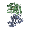
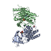
 集合体
集合体


 分子量: 62.005 Da / 分子数: 2 / 由来タイプ: 合成 / 式: NO3
 分子量: 18.015 Da / 分子数: 182 / 由来タイプ: 天然 / 式: H2O
 試料調製
試料調製 解析
解析