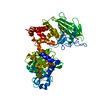
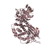
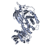
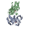
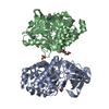
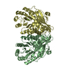
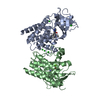
Entry Database : PDB / ID : 4bpcTitle Structure of the Catalytic Domain of Protein Tyrosine Phosphatase Sigma in the Sulfenic Acid Form RECEPTOR-TYPE TYROSINE-PROTEIN PHOSPHATASE S Keywords / / Function / homology Function Domain/homology Component
/ / / / / / / / / / / / / / / / / / / / / / / / / / / / / / / / / / / / / / / / / / / / / / / / / / / / / / / / / / / / / / / / / / / / / / / / / / / / / / / / / / / / / / / / / / Biological species HOMO SAPIENS (human)Method / / / Resolution : 2.1 Å Authors Jeon, T.J. / Chien, P.N. / Chun, H.J. / Ryu, S.E. Journal : Mol.Cells / Year : 2013Title : Structure of the Catalytic Domain of Protein Tyrosine Phosphatase Sigma in the Sulfenic Acid FormAuthors : Jeon, T.J. / Chien, P.N. / Chun, H.J. / Ryu, S.E. History Deposition May 24, 2013 Deposition site / Processing site Revision 1.0 Jul 17, 2013 Provider / Type Revision 1.1 Aug 7, 2013 Group Revision 1.2 Sep 23, 2015 Group Revision 1.3 Jan 30, 2019 Group / Experimental preparation / Category / Item Revision 1.4 Feb 6, 2019 Group / Experimental preparation / Category / Item Revision 1.5 Dec 20, 2023 Group Data collection / Database references ... Data collection / Database references / Derived calculations / Other / Refinement description Category chem_comp_atom / chem_comp_bond ... chem_comp_atom / chem_comp_bond / database_2 / pdbx_database_status / pdbx_initial_refinement_model / struct_conn Item _database_2.pdbx_DOI / _database_2.pdbx_database_accession ... _database_2.pdbx_DOI / _database_2.pdbx_database_accession / _pdbx_database_status.status_code_sf / _struct_conn.pdbx_leaving_atom_flag Revision 1.6 Oct 16, 2024 Group / Category / pdbx_modification_feature / Item
Show all Show less
 Movie
Movie Controller

 Yorodumi
 Open data
 Basic information
 Components
 Keywords
 Function and homology information
 HOMO SAPIENS (human)
 X-RAY DIFFRACTION /
  Authors
 Citation
 Structure visualization
 Downloads & links
 Other downloads
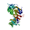


 PDBj
PDBj


 Assembly
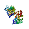

 Mass: 18.015 Da / Num. of mol.: 181 / Source method: isolated from a natural source / Formula: H2O
 Sample preparation
 / Beamline: 7A (6B, 6C1) / Wavelength: 1
  Processing