- PDB-2p4n: Human Monomeric Kinesin (1BG2) and Bovine Tubulin (1JFF) Docked i... -
+
データを開く
IDまたはキーワード:
読み込み中...
-
基本情報
登録情報
データベース: PDB / ID: 2p4n
タイトル
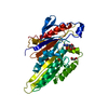
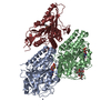
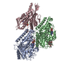
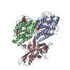
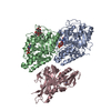
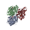
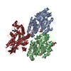
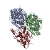
Human Monomeric Kinesin (1BG2) and Bovine Tubulin (1JFF) Docked into the 9-Angstrom Cryo-EM Map of Nucleotide-Free Kinesin Complexed to the Microtubule
要素
Kinesin heavy chain
Tubulin alpha chain
Tubulin beta chain
キーワード
TRANSPORT PROTEIN / Motor protein / ATPase
機能・相同性
機能・相同性情報
cytoplasm organization / cytolytic granule membrane / plus-end-directed vesicle transport along microtubule / mitocytosis / anterograde dendritic transport of neurotransmitter receptor complex / anterograde neuronal dense core vesicle transport / anterograde axonal protein transport / retrograde neuronal dense core vesicle transport / vesicle transport along microtubule / lysosome localization ...cytoplasm organization / cytolytic granule membrane / plus-end-directed vesicle transport along microtubule / mitocytosis / anterograde dendritic transport of neurotransmitter receptor complex / anterograde neuronal dense core vesicle transport / anterograde axonal protein transport / retrograde neuronal dense core vesicle transport / vesicle transport along microtubule / lysosome localization / positive regulation of potassium ion transport / natural killer cell mediated cytotoxicity / Kinesins / plus-end-directed microtubule motor activity / RHO GTPases activate KTN1 / stress granule disassembly / positive regulation of axon guidance / mitochondrion transport along microtubule / ciliary rootlet / centrosome localization / COPI-dependent Golgi-to-ER retrograde traffic / microtubule motor activity / kinesin complex / synaptic vesicle transport / Insulin processing / microtubule-based movement / microtubule-based process / centriolar satellite / axon cytoplasm / MHC class II antigen presentation / dendrite cytoplasm / phagocytic vesicle / regulation of membrane potential / positive regulation of synaptic transmission, GABAergic / positive regulation of protein localization to plasma membrane / 加水分解酵素; 酸無水物に作用; GTPに作用・細胞または細胞小器官の運動に関与 / axon guidance / structural constituent of cytoskeleton / cellular response to type II interferon / microtubule cytoskeleton organization / microtubule cytoskeleton / Signaling by ALK fusions and activated point mutants / nervous system development / mitotic cell cycle / microtubule binding / microtubule / vesicle / hydrolase activity / cadherin binding / protein heterodimerization activity / GTPase activity / protein-containing complex binding / GTP binding / perinuclear region of cytoplasm / ATP hydrolysis activity / mitochondrion / ATP binding / identical protein binding / membrane / metal ion binding / cytoplasm / cytosol 類似検索 - 分子機能
Kinesin-like protein / Kinesin motor domain signature. / Kinesin motor domain, conserved site / Kinesin motor domain / Kinesin motor domain profile. / Kinesin motor, catalytic domain. ATPase. / Kinesin motor domain / Kinesin motor domain superfamily / Alpha tubulin / Tubulin-beta mRNA autoregulation signal. ...Kinesin-like protein / Kinesin motor domain signature. / Kinesin motor domain, conserved site / Kinesin motor domain / Kinesin motor domain profile. / Kinesin motor, catalytic domain. ATPase. / Kinesin motor domain / Kinesin motor domain superfamily / Alpha tubulin / Tubulin-beta mRNA autoregulation signal. / Beta tubulin, autoregulation binding site / Beta tubulin / Tubulin / Tubulin, C-terminal / Tubulin C-terminal domain / Tubulin, conserved site / Tubulin subunits alpha, beta, and gamma signature. / Tubulin/FtsZ family, C-terminal domain / Tubulin/FtsZ-like, C-terminal domain / Tubulin/FtsZ, C-terminal / Tubulin/FtsZ, 2-layer sandwich domain / Tubulin/FtsZ family, GTPase domain / Tubulin/FtsZ family, GTPase domain / Tubulin/FtsZ, GTPase domain / Tubulin/FtsZ, GTPase domain superfamily / P-loop containing nucleoside triphosphate hydrolase 類似検索 - ドメイン・相同性
ジャーナル: J Cell Biol / 年: 2007 タイトル: The beginning of kinesin's force-generating cycle visualized at 9-A resolution. 著者: Charles V Sindelar / Kenneth H Downing / 要旨: We have used cryo-electron microscopy of kinesin-decorated microtubules to resolve the structure of the motor protein kinesin's crucial nucleotide response elements, switch I and the switch II helix, ...We have used cryo-electron microscopy of kinesin-decorated microtubules to resolve the structure of the motor protein kinesin's crucial nucleotide response elements, switch I and the switch II helix, in kinesin's poorly understood nucleotide-free state. Both of the switch elements undergo conformational change relative to the microtubule-free state. The changes in switch I suggest a role for it in "ejecting" adenosine diphosphate when kinesin initially binds to the microtubule. The switch II helix has an N-terminal extension, apparently stabilized by conserved microtubule contacts, implying a microtubule activation mechanism that could convey the state of the bound nucleotide to kinesin's putative force-delivering element (the "neck linker"). In deriving this structure, we have adapted an image-processing technique, single-particle reconstruction, for analyzing decorated microtubules. The resulting reconstruction visualizes the asymmetric seam present in native, 13-protofilament microtubules, and this method will provide an avenue to higher-resolution characterization of a variety of microtubule- binding proteins, as well as the microtubule itself.
Kinesinheavychain / Ubiquitous kinesin heavy chain / UKHC
分子量: 36405.070 Da / 分子数: 1 / 断片: K349 Construct of Human Kinesin / 由来タイプ: 天然 詳細: The actual construct used in the EM studies is a mutant protein (called cys-lite) 由来: (天然) Homo sapiens (ヒト) / 参照: UniProt: P33176*PLUS
THE SEQUENCE OF KINESIN (CHAIN K) IN THE SAMPLE INCLUDED MUTATIONS.THE CONSTRUCT IS KNOWN AS CYS- ...THE SEQUENCE OF KINESIN (CHAIN K) IN THE SAMPLE INCLUDED MUTATIONS.THE CONSTRUCT IS KNOWN AS CYS-LITE. MODEL CRYSTAL STRUCTURE FOR FITTING THE MAP WAS THE NATIVE SEQUENCE. IN THE CASE OF TUBULIN A AND B (CHAINS A AND B), THE SEQUENCE IN THE SAMPLE WAS DERIVED FROM COW, WHILE THE MODEL USED FOR FITTING THE MAP WAS A PIG PROTEIN.
-
実験情報
-
実験
実験
手法: 電子顕微鏡法
EM実験
試料の集合状態: FILAMENT / 3次元再構成法: 単粒子再構成法
-
試料調製
構成要素
ID
名称
タイプ
Parent-ID
詳細
1
Nucleotide-free kinesin bound to a 13-protofilament microtubule
COMPLEX
0
2
Kinesinheavychain
1
microtubuleassociatedcomplex
3
Tubulinalphachain
1
microtubule
4
Tubulinbetachain
1
microtubule
緩衝液
pH: 6.8
試料
包埋: YES / シャドウイング: NO / 染色: NO / 凍結: YES
試料支持
詳細: 300 mesh copper grid
急速凍結
装置: HOMEMADE PLUNGER / 凍結剤: ETHANE / 詳細: ETHANE
-
電子顕微鏡撮影
顕微鏡
モデル: JEOL 4000
電子銃
電子線源: LAB6 / 加速電圧: 400 kV / 照射モード: FLOOD BEAM
電子レンズ
モード: BRIGHT FIELD / 倍率(公称値): 60000 X / 最大 デフォーカス(公称値): 1500 nm / 最小 デフォーカス(公称値): 700 nm / Cs: 4.1 mm
試料ホルダ
温度: 103.15 K / 傾斜角・最大: 0 ° / 傾斜角・最小: 0 °
撮影
電子線照射量: 16 e/Å2 / フィルム・検出器のモデル: KODAK SO-163 FILM
-
解析
EMソフトウェア
ID
名称
カテゴリ
1
Situs
モデルフィッティング
2
SPIDER
3次元再構成
CTF補正
詳細: CTF correction was integrated into the back projection process with a customized C program
3次元再構成
手法: Back projection with integrated CTF correction / 解像度: 9 Å / 粒子像の数: 150000 / ピクセルサイズ(公称値): 1 Å / ピクセルサイズ(実測値): 0.98 Å 倍率補正: The magnification was calibrated by assuming a microtubule dimer spacing of 80.0 Angstroms. 詳細: Single-particle analysis was employed. / 対称性のタイプ: HELICAL
 ムービー
ムービー コントローラー


 データを開く
 基本情報
基本情報 要素
要素 キーワード
キーワード 機能・相同性情報
 Homo sapiens (ヒト)
 データ登録者
 引用
引用
 構造の表示
構造の表示 UCSF Chimera
 ダウンロードとリンク
 その他のダウンロード
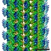


 PDBj
PDBj


 集合体
集合体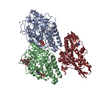


 分子量: 24.305 Da / 分子数: 2 / 由来タイプ: 合成 / 式: Mg
 分子量: 427.201 Da / 分子数: 1 / 由来タイプ: 合成 / 式: C10H15N5O10P2 / コメント: ADP, エネルギー貯蔵分子*YM
 分子量: 65.409 Da / 分子数: 1 / 由来タイプ: 合成 / 式: Zn
 分子量: 523.180 Da / 分子数: 1 / 由来タイプ: 合成 / 式: C10H16N5O14P3 / コメント: GTP, エネルギー貯蔵分子*YM
 タイプ: RNA linking / 分子量: 443.201 Da / 分子数: 1 / 由来タイプ: 合成 / 式: C10H15N5O11P2 / コメント: GDP, エネルギー貯蔵分子*YM
 分子量: 853.906 Da / 分子数: 1 / 由来タイプ: 合成 / 式: C47H51NO14 / コメント: 薬剤, 化学療法薬*YM
 試料調製
試料調製 電子顕微鏡撮影
 解析
解析