 Movie
Movie Controller


+ Open data

- Basic information
| Entry | Database: PDB / ID: 2oz9 | |||||||||
|---|---|---|---|---|---|---|---|---|---|---|
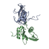
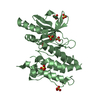
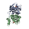
| Title | E. coli TRP holorepressor, orthorhombic crystal form | |||||||||
 Components Components | Trp operon repressor | |||||||||
 Keywords Keywords | DNA BINDING PROTEIN / DNA BINDING REGULATORY PROTEIN | |||||||||
| Function / homology |  Function and homology information Function and homology informationsequence-specific DNA binding / DNA-binding transcription factor activity / negative regulation of DNA-templated transcription / regulation of DNA-templated transcription / DNA binding / cytoplasm Similarity search - Function | |||||||||
| Biological species |  | |||||||||
| Method |  X-RAY DIFFRACTION / MOLECULAR REPLACEMENT / Resolution: 1.65 Å X-RAY DIFFRACTION / MOLECULAR REPLACEMENT / Resolution: 1.65 Å | |||||||||
 Authors Authors | Lawson, C.L. / Sigler, P.B. | |||||||||
 Citation Citation | Journal: Proteins / Year: 1988 Title: Flexibility of the DNA-binding domains of trp repressor. Authors: Lawson, C.L. / Zhang, R.G. / Schevitz, R.W. / Otwinowski, Z. / Joachimiak, A. / Sigler, P.B. #1: Journal: Nature / Year: 1987Title: The Crystal Structure of Trp Aporepressor at 1.8 Angstroms Shows How Binding Tryptophan Enhances DNA Affinity Authors: Zhang, R.G. / Joachimiak, A. / Lawson, C.L. / Schevitz, R.W. / Otwinowski, Z. / Sigler, P.B. #2: Journal: Nature / Year: 1985Title: The Three-Dimensional Structure of Trp Repressor Authors: Schevitz, R.W. / Otwinowski, Z. / Joachimiak, A. / Lawson, C.L. / Sigler, P.B. #3: Journal: J.Biol.Chem. / Year: 1983Title: Functional Inferences from Crystals of Escherichia Coli Trp Repressor Authors: Joachimiak, A. / Schevitz, R.W. / Kelley, R.L. / Yanofsky, C. / Sigler, P.B. #4: Journal: Proc.Natl.Acad.Sci.USA / Year: 1983Title: Purification and Characterization of Trp Repressor Authors: Joachimiak, A. / Kelley, R.L. / Gunsalus, R.P. / Yanofsky, C. / Sigler, P.B. #5: Journal: Proc.Natl.Acad.Sci.USA / Year: 1980Title: Nucleotide Sequence and Expression of Escherichia Coli Trpr, the Structural Gene for the Trp Aporepressor Authors: Gunsalus, R.P. / Yanofsky, C. | |||||||||
| History |
|
- Structure visualization
| Structure viewer | Molecule: MolmilJmol/JSmol |
|---|
- Downloads & links
-Download
| PDBx/mmCIF format | 2oz9.cif.gz | 37.4 KB | Display | PDBx/mmCIF format |
|---|---|---|---|---|
| PDB format | pdb2oz9.ent.gz | 24.4 KB | Display | PDB format |
| PDBx/mmJSON format | 2oz9.json.gz | Tree view | PDBx/mmJSON format | |
| Others |  Other downloads Other downloads |
-Validation report
| Arichive directory | https://data.pdbj.org/pub/pdb/validation_reports/oz/2oz9ftp://data.pdbj.org/pub/pdb/validation_reports/oz/2oz9 | HTTPS FTP |
|---|
-Related structure data
| Related structure data | 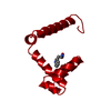 1wrpSC  3wrpC S: Starting model for refinement C: citing same article ( |
|---|---|
| Similar structure data |


-Links
 PDBj
PDBj

- Assembly
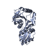
| Deposited unit | 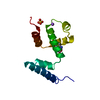
| ||||||||
|---|---|---|---|---|---|---|---|---|---|
| 1 | 
| ||||||||
| Unit cell |
| ||||||||
| Details | The biological assembly is a dimer. To generate the second half of the assembly apply the crystallographic symmetry operator -x, -y, z. |
-Components
| #1: Protein | Mass: 12238.934 Da / Num. of mol.: 1 Source method: isolated from a genetically manipulated source Source: (gene. exp.) |
|---|---|
| #2: Chemical | ChemComp-NA /   Mass: 22.990 Da / Num. of mol.: 1 / Source method: obtained synthetically / Formula: Na Mass: 22.990 Da / Num. of mol.: 1 / Source method: obtained synthetically / Formula: Na |
| #3: Chemical | ChemComp-SO4 /   Mass: 96.063 Da / Num. of mol.: 1 / Source method: obtained synthetically / Formula: SO4 Mass: 96.063 Da / Num. of mol.: 1 / Source method: obtained synthetically / Formula: SO4 |
| #4: Chemical | ChemComp-TRP /   Type: L-peptide linking / Mass: 204.225 Da / Num. of mol.: 1 / Source method: obtained synthetically / Formula: C11H12N2O2 Type: L-peptide linking / Mass: 204.225 Da / Num. of mol.: 1 / Source method: obtained synthetically / Formula: C11H12N2O2 |
| #5: Water | ChemComp-HOH /  Mass: 18.015 Da / Num. of mol.: 85 / Source method: isolated from a natural source / Formula: H2O Mass: 18.015 Da / Num. of mol.: 85 / Source method: isolated from a natural source / Formula: H2O |
-Experimental details
-Experiment
| Experiment | Method: X-RAY DIFFRACTION / Number of used crystals: 1 |
|---|
- Sample preparation
| Crystal | Density Matthews: 1.93 Å3/Da / Density % sol: 36.3 % |
|---|---|
| Crystal grow | Temperature: 298 K / Method: vapor diffusion / pH: 5 Details: crystallization conditions: 2.5 M sodium phosphate, 0.6 M ammonium chloride, 2.5 mM L-tryptophan. Prior to data collection the crystal was equilibrated to 2.4 M ammonium sulfate, 0.4 M ...Details: crystallization conditions: 2.5 M sodium phosphate, 0.6 M ammonium chloride, 2.5 mM L-tryptophan. Prior to data collection the crystal was equilibrated to 2.4 M ammonium sulfate, 0.4 M sodium chloride, 2.4 mM L-tryptophan, 50 mM sodium acetate., pH 5.0, VAPOR DIFFUSION, temperature 298K |
-Data collection
| Diffraction | Mean temperature: 298 K |
|---|---|
| Diffraction source | Source: ROTATING ANODE / Type: ELLIOTT GX-6 / Wavelength: 1.5418 Å |
| Detector | Type: KODAK / Detector: FILM / Details: graphite monochromator |
| Radiation | Monochromator: graphite / Protocol: SINGLE WAVELENGTH / Monochromatic (M) / Laue (L): M / Scattering type: x-ray |
| Radiation wavelength | Wavelength: 1.5418 Å / Relative weight: 1 |
| Reflection | Resolution: 1.65→20 Å / Num. all: 10945 / Num. obs: 10945 / Observed criterion σ(F): 2 |
- Processing
| Software |
| ||||||||||||
|---|---|---|---|---|---|---|---|---|---|---|---|---|---|
| Refinement | Method to determine structure: MOLECULAR REPLACEMENT Starting model: 1wrp dimer Resolution: 1.65→5 Å / Isotropic thermal model: isotropic / Cross valid method: NONE / σ(F): 2 / Stereochemistry target values: Hendrickson & Konnert Details: REFINEMENT. BY THE RESTRAINED LEAST-SQUARES PROCEDURE OF J. KONNERT AND W. HENDRICKSON (PROGRAM *PROLSQ*) AS MODIFIED BY B. FINZEL (PROGRAM *PROFFT*) AND USE OF PROGRAM *FRODO* OF T. A. ...Details: REFINEMENT. BY THE RESTRAINED LEAST-SQUARES PROCEDURE OF J. KONNERT AND W. HENDRICKSON (PROGRAM *PROLSQ*) AS MODIFIED BY B. FINZEL (PROGRAM *PROFFT*) AND USE OF PROGRAM *FRODO* OF T. A. JONES. THE R VALUE IS 0.180. THE RMS DEVIATION FROM IDEALITY OF THE BOND LENGTHS IS 0.012 ANGSTROMS. THE RMS DEVIATION FROM IDEALITY OF THE BOND ANGLES IS 2.0 DEGREES.
| ||||||||||||
| Displacement parameters | Biso mean: 17.284 Å2 | ||||||||||||
| Refine analyze | Luzzati coordinate error obs: 0.2 Å | ||||||||||||
| Refinement step | Cycle: LAST / Resolution: 1.65→5 Å
| ||||||||||||
| Refine LS restraints |
|
