 Movie
Movie Controller


+ Open data

- Basic information
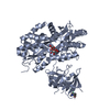
| Entry | Database: PDB / ID: 2hur | ||||||
|---|---|---|---|---|---|---|---|
| Title | Escherichia coli nucleoside diphosphate kinase | ||||||
 Components Components | NUCLEOSIDE DIPHOSPHATE KINASE | ||||||
 Keywords Keywords | SIGNALING PROTEIN / TRANSFERASE / TYPE II TETRAMER | ||||||
| Function / homology |  Function and homology information Function and homology informationpurine nucleotide metabolic process / pyrimidine nucleotide metabolic process / nucleoside-diphosphate kinase / UTP biosynthetic process / CTP biosynthetic process / nucleoside diphosphate kinase activity / GTP biosynthetic process / ATP binding / metal ion binding / cytosol Similarity search - Function | ||||||
| Biological species |  | ||||||
| Method |  X-RAY DIFFRACTION / SYNCHROTRON / MOLECULAR REPLACEMENT / Resolution: 1.62 Å X-RAY DIFFRACTION / SYNCHROTRON / MOLECULAR REPLACEMENT / Resolution: 1.62 Å | ||||||
 Authors Authors | Moynie, L. / Giraud, M.-F. / Georgescauld, F. / Lascu, I. / Dautant, A. | ||||||
 Citation Citation | Journal: Proteins / Year: 2007 Title: The structure of the Escherichia coli nucleoside diphosphate kinase reveals a new quaternary architecture for this enzyme family Authors: Moynie, L. / Giraud, M.-F. / Georgescauld, F. / Lascu, I. / Dautant, A. #1: Journal: J.Bacteriol. / Year: 1995 Title: Nucleoside diphosphate kinase from Escherichia coli Authors: Almaula, N. / Lu, Q. / Delgado, J. / Belkin, S. / Inouye, M. #2: Journal: J.Mol.Biol. / Year: 1993Title: Crystal structure of Myxococcus xanthus nucleoside diphosphate kinase and its interaction with a nucleotide substrate at 2.0 A resolution Authors: Williams, R.L. / Oren, D.A. / Inouye, S. / Inouye, M. / Arnold, E. #3: Journal: J.Mol.Biol. / Year: 1994Title: Refined X-ray structure of Dictyostelium discoideum nucleoside diphosphate kinase at 1.8 A resolution. Authors: LeBras, G. / Lascu, I. / Lacombe, M.L. / Janin, J. | ||||||
| History |
|
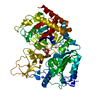
- Structure visualization
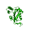
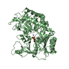
| Structure viewer | Molecule: MolmilJmol/JSmol |
|---|
- Downloads & links
-Download
| PDBx/mmCIF format | 2hur.cif.gz | 183.4 KB | Display | PDBx/mmCIF format |
|---|---|---|---|---|
| PDB format | pdb2hur.ent.gz | 146.4 KB | Display | PDB format |
| PDBx/mmJSON format | 2hur.json.gz | Tree view | PDBx/mmJSON format | |
| Others |  Other downloads Other downloads |
-Validation report
| Arichive directory | https://data.pdbj.org/pub/pdb/validation_reports/hu/2hurftp://data.pdbj.org/pub/pdb/validation_reports/hu/2hur | HTTPS FTP |
|---|
-Related structure data
| Related structure data | 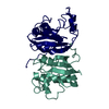 2nckS S: Starting model for refinement |
|---|---|
| Similar structure data |


-Links
 PDBj
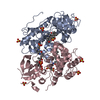
PDBj- Assembly
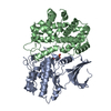
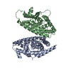
| Deposited unit | 
| ||||||||||||||||||||||||
|---|---|---|---|---|---|---|---|---|---|---|---|---|---|---|---|---|---|---|---|---|---|---|---|---|---|
| 1 | 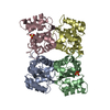
| ||||||||||||||||||||||||
| 2 | 
| ||||||||||||||||||||||||
| 3 | 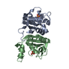
| ||||||||||||||||||||||||
| 4 | 
| ||||||||||||||||||||||||
| 5 | 
| ||||||||||||||||||||||||
| Unit cell |
| ||||||||||||||||||||||||
| Components on special symmetry positions |
|
-Components
| #1: Protein | Mass: 15350.208 Da / Num. of mol.: 6 Source method: isolated from a genetically manipulated source Source: (gene. exp.) #2: Chemical | ChemComp-SO4 /   Mass: 96.063 Da / Num. of mol.: 6 / Source method: obtained synthetically / Formula: SO4 Mass: 96.063 Da / Num. of mol.: 6 / Source method: obtained synthetically / Formula: SO4#3: Water | ChemComp-HOH / |  Mass: 18.015 Da / Num. of mol.: 717 / Source method: isolated from a natural source / Formula: H2O Mass: 18.015 Da / Num. of mol.: 717 / Source method: isolated from a natural source / Formula: H2O |
|---|
-Experimental details
-Experiment
| Experiment | Method: X-RAY DIFFRACTION / Number of used crystals: 1 |
|---|
- Sample preparation
| Crystal | Density Matthews: 2.34 Å3/Da / Density % sol: 47.5 % |
|---|---|
| Crystal grow | Temperature: 293 K / Method: vapor diffusion, sitting drop / pH: 4.6 Details: 0.1 M AMMONIUM SULPHATE, 25% PEG 4000 0.1 M SODIUM ACETATE/ACETIC ACID, pH 4.6, VAPOR DIFFUSION, SITTING DROP, temperature 293K |
-Data collection
| Diffraction | Mean temperature: 107 K |
|---|---|
| Diffraction source | Source: SYNCHROTRON / Site: ESRF  / Beamline: ID14-1 / Wavelength: 0.934 / Wavelength: 0.934 Å / Beamline: ID14-1 / Wavelength: 0.934 / Wavelength: 0.934 Å |
| Detector | Type: ADSC QUANTUM 4 / Detector: CCD / Date: Feb 24, 2006 |
| Radiation | Protocol: SINGLE WAVELENGTH / Monochromatic (M) / Laue (L): M / Scattering type: x-ray |
| Radiation wavelength | Wavelength: 0.934 Å / Relative weight: 1 |
| Reflection | Resolution: 1.62→38.05 Å / Num. all: 108681 / Num. obs: 108681 / % possible obs: 99.7 % / Observed criterion σ(F): 0 / Observed criterion σ(I): 0 / Redundancy: 3.5 % / Biso Wilson estimate: 19.5 Å2 / Rsym value: 0.09 / Net I/σ(I): 16.6 |
| Reflection shell | Resolution: 1.62→1.66 Å / Redundancy: 2.8 % / Mean I/σ(I) obs: 2.1 / Rsym value: 0.38 / % possible all: 97.1 |
- Processing
| Software |
| ||||||||||||||||||||||||||||||
|---|---|---|---|---|---|---|---|---|---|---|---|---|---|---|---|---|---|---|---|---|---|---|---|---|---|---|---|---|---|---|---|
| Refinement | Method to determine structure: MOLECULAR REPLACEMENT Starting model: PDB FILE 2NCK Resolution: 1.62→38.05 Å / Rfactor Rfree error: 0.003 / Data cutoff high absF: 2343463.17 / Data cutoff low absF: 0 / Isotropic thermal model: RESTRAINED / Cross valid method: THROUGHOUT / σ(F): 0 / σ(I): 0
| ||||||||||||||||||||||||||||||
| Solvent computation | Solvent model: FLAT MODEL / Bsol: 45.0217 Å2 / ksol: 0.368627 e/Å3 | ||||||||||||||||||||||||||||||
| Displacement parameters | Biso mean: 21.4 Å2
| ||||||||||||||||||||||||||||||
| Refine analyze |
| ||||||||||||||||||||||||||||||
| Refinement step | Cycle: LAST / Resolution: 1.62→38.05 Å
| ||||||||||||||||||||||||||||||
| Refine LS restraints |
| ||||||||||||||||||||||||||||||
| LS refinement shell | Resolution: 1.62→1.72 Å / Rfactor Rfree error: 0.01 / Total num. of bins used: 6
| ||||||||||||||||||||||||||||||
| Xplor file |
|
