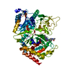
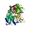
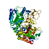
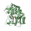
Entry Database : PDB / ID : 2gmvTitle PEPCK complex with a GTP-competitive inhibitor Phosphoenolpyruvate carboxykinase, cytosolic Keywords / / / Function / homology Function Domain/homology Component
/ / / / / / / / / / / / / / / / / / / / / / / / / / / / / / / / / / / / / / / / / / / / / / / / / / / / / / / / / / / / / / / / / / / / / / / / / / / / Biological species Homo sapiens (human)Method / / / Resolution : 2.3 Å Authors Dunten, P. Journal : Bioorg.Med.Chem.Lett. / Year : 2007Title : C-8 Modifications of 3-alkyl-1,8-dibenzylxanthines as inhibitors of human cytosolic phosphoenolpyruvate carboxykinase.
Authors :
Pietranico, S.L. / Foley, L.H. / Huby, N. / Yun, W. / Dunten, P. / Vermeulen, J. / Wang, P. / Toth, K. / Ramsey, G. / Gubler, M.L. / Wertheimer, S.J. #2: Journal : J.Mol.Biol. / Year : 2002Title : Crystal structure of human cytosolic phosphoenolpyruvate carboxykinase reveals a new GTP-binding site.
Authors :
Dunten, P. / Belunis, C. / Crowther, R. / Hollfelder, K. / Kammlott, U. / Levin, W. / Hanspeter, M. / Ramsey, G.B. / Swain, A. / Weber, D. / Wertheimer, S.J. History Deposition Apr 7, 2006 Deposition site / Processing site Revision 1.0 May 29, 2007 Provider / Type Revision 1.1 May 1, 2008 Group Revision 1.2 Jul 13, 2011 Group Revision 1.3 Jul 24, 2019 Group / Refinement description / Category / Item / _software.versionRevision 1.4 Feb 14, 2024 Group / Database references / Derived calculationsCategory chem_comp_atom / chem_comp_bond ... chem_comp_atom / chem_comp_bond / database_2 / pdbx_struct_conn_angle / struct_conn / struct_ref_seq_dif / struct_site Item _database_2.pdbx_DOI / _database_2.pdbx_database_accession ... _database_2.pdbx_DOI / _database_2.pdbx_database_accession / _pdbx_struct_conn_angle.ptnr1_auth_comp_id / _pdbx_struct_conn_angle.ptnr1_auth_seq_id / _pdbx_struct_conn_angle.ptnr1_label_atom_id / _pdbx_struct_conn_angle.ptnr1_label_comp_id / _pdbx_struct_conn_angle.ptnr1_label_seq_id / _pdbx_struct_conn_angle.ptnr3_auth_comp_id / _pdbx_struct_conn_angle.ptnr3_auth_seq_id / _pdbx_struct_conn_angle.ptnr3_label_atom_id / _pdbx_struct_conn_angle.ptnr3_label_comp_id / _pdbx_struct_conn_angle.ptnr3_label_seq_id / _pdbx_struct_conn_angle.value / _struct_conn.pdbx_dist_value / _struct_conn.ptnr1_auth_comp_id / _struct_conn.ptnr1_auth_seq_id / _struct_conn.ptnr1_label_asym_id / _struct_conn.ptnr1_label_atom_id / _struct_conn.ptnr1_label_comp_id / _struct_conn.ptnr1_label_seq_id / _struct_conn.ptnr2_auth_comp_id / _struct_conn.ptnr2_auth_seq_id / _struct_conn.ptnr2_label_asym_id / _struct_conn.ptnr2_label_atom_id / _struct_conn.ptnr2_label_comp_id / _struct_conn.ptnr2_label_seq_id / _struct_ref_seq_dif.details / _struct_site.pdbx_auth_asym_id / _struct_site.pdbx_auth_comp_id / _struct_site.pdbx_auth_seq_id
Show all Show less
 Movie
Movie Controller


 Open data
 Basic information
 Components
 Keywords
 Function and homology information
 Homo sapiens (human)
 X-RAY DIFFRACTION /
  Authors
 Citation
 Structure visualization
 Downloads & links
 Other downloads
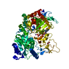


 PDBj
PDBj


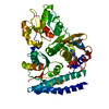
 Assembly
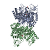


 Mass: 54.938 Da / Num. of mol.: 2 / Source method: obtained synthetically / Formula: Mn
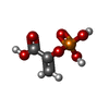
 Mass: 168.042 Da / Num. of mol.: 2 / Source method: obtained synthetically / Formula: C3H5O6P
 Mass: 565.619 Da / Num. of mol.: 1 / Source method: obtained synthetically / Formula: C27H28FN7O4S
 Mass: 18.015 Da / Num. of mol.: 132 / Source method: isolated from a natural source / Formula: H2O
 Sample preparation
 / Beamline: X8C
 Processing