[English] 日本語
Yorodumi
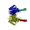
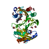
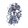
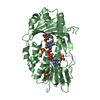
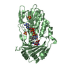
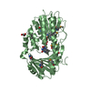
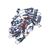
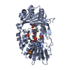
- PDB-2ovi: Structure of the Heme Binding Protein ChuX -

+
Open data


ID or keywords:

Loading...

-
Basic information

Entry
Database: PDB / ID: 2ovi
TitleStructure of the Heme Binding Protein ChuX
ComponentsHypothetical protein chuXHypothesis
KeywordsLIGAND BINDING PROTEIN / METAL TRANSPORT / 2 sets of 9 antiparallel beta sheet core flanked by 2 sets of 3 helices and another 2 sets of helices / Structural Genomics / Montreal-Kingston Bacterial Structural Genomics Initiative / BSGI
Function / homologyIntracellular heme transport protein HutX-like / Haem utilisation ChuX/HutX / HemS/ChuS/ChuX like domains / Heme iron utilization protein-like fold / 3-Layer(aba) Sandwich / Alpha Beta / Heme utilization carrier protein
Function and homology information
Biological speciesEscherichia coli O157:H7 (bacteria)
MethodX-RAY DIFFRACTION / SYNCHROTRON / SAD / Resolution: 2.05 Å
AuthorsSuits, M.D.L. / Pal, G.P. / Jia, Z. / Montreal-Kingston Bacterial Structural Genomics Initiative (BSGI)
CitationJournal: Protein Sci. / Year: 2009
Title: Structure and heme binding properties of Escherichia coli O157:H7 ChuX.
Authors: Suits, M.D. / Lang, J. / Pal, G.P. / Couture, M. / Jia, Z.
History
DepositionFeb 13, 2007Deposition site: RCSB / Processing site: RCSB
Revision 1.0Feb 12, 2008Provider: repository / Type: Initial release
Revision 1.1Jul 13, 2011Group: Version format compliance
Revision 1.2Oct 18, 2017Group: Refinement description / Category: software
Item: _software.classification / _software.contact_author ..._software.classification / _software.contact_author / _software.contact_author_email / _software.date / _software.language / _software.location / _software.name / _software.type / _software.version
Revision 1.3Oct 25, 2017Group: Author supporting evidence / Category: pdbx_struct_assembly_auth_evidence
Revision 1.4Feb 21, 2024Group: Data collection / Database references / Category: chem_comp_atom / chem_comp_bond / database_2
Item: _database_2.pdbx_DOI / _database_2.pdbx_database_accession
Remark 300 
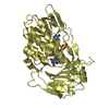
BIOMOLECULE: 1 THIS ENTRY CONTAINS THE CRYSTALLOGRAPHIC ASYMMETRIC UNIT WHICH CONSISTS OF 4 ... BIOMOLECULE: 1 THIS ENTRY CONTAINS THE CRYSTALLOGRAPHIC ASYMMETRIC UNIT WHICH CONSISTS OF 4 CHAIN(S). AUTHORS STATE THAT THE BIOLOGICAL ASSEMBLY IS A PAIR OF CHUX HOMODIMERS, SUGGESTED FROM THE CRYSTAL STRUCTURE AND SIZE EXCLUSION CHROMATOGRAPHY. SEE REMARK 350 FOR INFORMATION ON GENERATING THE BIOLOGICAL MOLECULE(S).

-
Structure visualization

Structure viewerMolecule:
MolmilJmol/JSmol

Downloads & links

-
Assembly

Deposited unit
A: Hypothetical protein chuX
B: Hypothetical protein chuX
C: Hypothetical protein chuX
D: Hypothetical protein chuX


Theoretical massNumber of molelcules
Total (without water)73,4114
Polymers73,4114
Non-polymers00
Water6,485360
1
A: Hypothetical protein chuX
D: Hypothetical protein chuX


Theoretical massNumber of molelcules
Total (without water)36,7062
Polymers36,7062
Non-polymers00
Water362
TypeNameSymmetry operationNumber
identity operation1_555x,y,z1
2
B: Hypothetical protein chuX
C: Hypothetical protein chuX


Theoretical massNumber of molelcules
Total (without water)36,7062
Polymers36,7062
Non-polymers00
Water362
TypeNameSymmetry operationNumber
identity operation1_555x,y,z1
Unit cell
Length a, b, c (Å)76.560, 76.560, 140.864
Angle α, β, γ (deg.)90.000, 90.000, 90.000
Int Tables number76
Space group name H-MP41
DetailsThe biological assembly is a pair of ChuX homodimers, suggested from the crystal structure and size exclusion chromatography

-
Components

#1: Protein
Hypothetical protein chuX / Hypothesis / ShuX-like protein


Mass: 18352.816 Da / Num. of mol.: 4
Source method: isolated from a genetically manipulated source
Source: (gene. exp.) Escherichia coli O157:H7 (bacteria) / Species: Escherichia coli / Strain: O157:H7, EDL933, EHEC / Gene: chuX, ECs4384, Z4915 / Plasmid: pET15b / Species (production host): Escherichia coli / Production host: Escherichia coli BL21(DE3) (bacteria) / Strain (production host): BL21(DE3) / References: UniProt: Q8X5N5
#2: Water ChemComp-HOH / water / Water


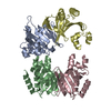
Mass: 18.015 Da / Num. of mol.: 360 / Source method: isolated from a natural source / Formula: H2O

-
Experimental details

-
Experiment

ExperimentMethod: X-RAY DIFFRACTION / Number of used crystals: 1

-
Sample preparation

CrystalDensity Matthews: 2.81 Å3/Da / Density % sol: 56.25 %
Crystal growTemperature: 295 K / Method: vapor diffusion, hanging drop / pH: 7.5
Details: 40 mg/ml protein, equal amount of protein solution and reservoir solution consisting of 1.50 M Ammonium sulfate, 50mM Hepes, pH 7.5, 2% (v/v) PEG 400, crystal appeared after 2-4 days, ...Details: 40 mg/ml protein, equal amount of protein solution and reservoir solution consisting of 1.50 M Ammonium sulfate, 50mM Hepes, pH 7.5, 2% (v/v) PEG 400, crystal appeared after 2-4 days, cryoprotection: Paratone-N oil, VAPOR DIFFUSION, HANGING DROP, temperature 295K

-
Data collection

Diffraction
IDMean temperature (K)Crystal-ID
11001
21001
Diffraction source
SourceSiteBeamlineIDWavelength (Å)
SYNCHROTRONNSLS X8C10.9795
SYNCHROTRONNSLS X12C21.1
Detector
TypeIDDetectorDate
ADSC QUANTUM 3151CCDApr 15, 2005
ADSC QUANTUM 3152CCDApr 27, 2006
Radiation
IDMonochromatorProtocolMonochromatic (M) / Laue (L)Scattering typeWavelength-ID
1Si(111)SINGLE WAVELENGTHMx-ray1
2Si(111)SINGLE WAVELENGTHMx-ray1
Radiation wavelength
IDWavelength (Å)Relative weight
10.97951
21.11
ReflectionResolution: 2.05→50 Å / Num. obs: 50833 / % possible obs: 100 % / Redundancy: 7.1 % / Rmerge(I) obs: 0.055 / Χ2: 0.957 / Net I/σ(I): 15.1
Reflection shellResolution: 2.05→2.12 Å / Redundancy: 6.9 % / Rmerge(I) obs: 0.95 / Num. unique all: 5077 / Χ2: 0.701 / % possible all: 100

-
Phasing

PhasingMethod: SAD
Phasing set
ID
1
2
3
Phasing MAD set
Clust-IDExpt-IDSet-IDWavelength (Å)F double prime refinedF prime refined
13 wavelength10.97950.47-2.21
13 wavelength20.97970.82-14.27
13 wavelength31.10.63-2.23
Phasing dmFOM : 0.48 / FOM acentric: 0.47 / FOM centric: 0.5 / Reflection: 35240 / Reflection acentric: 34324 / Reflection centric: 916
Phasing dm shell
Resolution (Å)FOM FOM acentricFOM centricReflectionReflection acentricReflection centric
6.3-19.9310.640.660.4915811476105
3.9-6.30.490.490.4849584777181
3.1-3.90.460.450.662356067168
2.8-3.10.480.480.6162496109140
2.4-2.80.510.510.531110910898211
2.2-2.40.350.350.2151084997111

-
Processing

Software
NameVersionClassificationNB
DENZOdata reduction
SCALEPACKdata scaling
SOLVE2.06phasing
RESOLVE2.06phasing
CNSrefinement
PDB_EXTRACT2data extraction
ADSCQuantumdata collection
HKL-2000data reduction
RefinementMethod to determine structure: SAD / Resolution: 2.05→50 Å / FOM work R set: 0.837 / σ(F): 0
RfactorNum. reflection% reflection
Rfree0.25 3137 6.2 %
Rwork0.213 --
obs-47915 94.4 %
Solvent computationBsol: 72.994 Å2
Displacement parametersBiso mean: 49.899 Å2
Baniso -1Baniso -2Baniso -3
1--3.216 Å20 Å20 Å2
2---3.216 Å20 Å2
3---6.432 Å2
Refinement stepCycle: LAST / Resolution: 2.05→50 Å
ProteinNucleic acidLigandSolventTotal
Num. atoms4832 0 0 360 5192
Refine LS restraints
Refine-IDTypeDev idealDev ideal target
X-RAY DIFFRACTIONc_mcbond_it4.3311.5
X-RAY DIFFRACTIONc_scbond_it6.6362
X-RAY DIFFRACTIONc_mcangle_it5.5772
X-RAY DIFFRACTIONc_scangle_it8.3952.5
LS refinement shell

Refine-ID: X-RAY DIFFRACTION / Total num. of bins used: 50

Resolution (Å)Rfactor RfreeNum. reflection RfreeRfactor RworkNum. reflection RworkNum. reflection obs
2.05-2.060.279650.293780845
2.06-2.080.346590.291773832
2.08-2.090.298590.304784843
2.09-2.110.38570.292798855
2.11-2.120.298490.287813862
2.12-2.140.27530.266833886
2.14-2.160.278400.28769809
2.16-2.170.292650.269870935
2.17-2.190.325620.266847909
2.19-2.210.284490.242822871
2.21-2.230.254620.219843905
2.23-2.250.291610.236881942
2.25-2.270.295590.258838897
2.27-2.290.301620.248886948
2.29-2.310.296540.241908962
2.31-2.330.3640.232863927
2.33-2.350.288480.225894942
2.35-2.380.27750.224831906
2.38-2.40.305570.229915972
2.4-2.430.312660.232859925
2.43-2.460.268670.226907974
2.46-2.490.27660.219874940
2.49-2.520.268770.233901978
2.52-2.550.232500.218930980
2.55-2.580.377600.229896956
2.58-2.620.23570.183897954
2.62-2.660.327660.2269391005
2.66-2.70.25620.21908970
2.7-2.740.312700.229381008
2.74-2.780.247640.212892956
2.78-2.830.31710.2369351006
2.83-2.880.269730.227914987
2.88-2.940.357790.2349221001
2.94-30.241670.228923990
3-3.060.288650.2249601025
3.06-3.130.321630.234898961
3.13-3.210.252610.2359701031
3.21-3.30.315670.2159441011
3.3-3.40.286590.21936995
3.4-3.510.231630.2139541017
3.51-3.630.264720.2179481020
3.63-3.780.234660.1949391005
3.78-3.950.209570.1959431000
3.95-4.160.207770.1889531030
4.16-4.420.152680.1589431011
4.42-4.760.207660.1569651031
4.76-5.230.21610.1799501011
5.23-5.990.268650.229661031
5.99-7.540.231610.2469561017
7.54-500.207710.2139701041
Xplor file
Refine-IDSerial noParam file
X-RAY DIFFRACTION1CNS_TOPPAR:protein_rep.param
X-RAY DIFFRACTION2CNS_TOPPAR:water_rep.param
X-RAY DIFFRACTION3CNS_TOPPAR:ion.param
X-RAY DIFFRACTION4hdm_xplor_par.txt

+
About Yorodumi

-
News

-
Feb 9, 2022. New format data for meta-information of EMDB entries

New format data for meta-information of EMDB entries

  • Version 3 of the EMDB header file is now the official format.
  • The previous official version 1.9 will be removed from the archive.

Related info.:EMDB header

External links:wwPDB to switch to version 3 of the EMDB data model

-
Aug 12, 2020. Covid-19 info

Covid-19 info

URL: https://pdbj.org/emnavi/covid19.php

New page: Covid-19 featured information page in EM Navigator.

Related info.:Covid-19 info / Mar 5, 2020. Novel coronavirus structure data

+
Mar 5, 2020. Novel coronavirus structure data

Novel coronavirus structure data

Related info.:Yorodumi Speices / Aug 12, 2020. Covid-19 info

External links:COVID-19 featured content - PDBj / Molecule of the Month (242):Coronavirus Proteases

+
Jan 31, 2019. EMDB accession codes are about to change! (news from PDBe EMDB page)

EMDB accession codes are about to change! (news from PDBe EMDB page)

  • The allocation of 4 digits for EMDB accession codes will soon come to an end. Whilst these codes will remain in use, new EMDB accession codes will include an additional digit and will expand incrementally as the available range of codes is exhausted. The current 4-digit format prefixed with “EMD-” (i.e. EMD-XXXX) will advance to a 5-digit format (i.e. EMD-XXXXX), and so on. It is currently estimated that the 4-digit codes will be depleted around Spring 2019, at which point the 5-digit format will come into force.
  • The EM Navigator/Yorodumi systems omit the EMD- prefix.

Related info.:Q: What is EMD? / ID/Accession-code notation in Yorodumi/EM Navigator

External links:EMDB Accession Codes are Changing Soon! / Contact to PDBj

+
Jul 12, 2017. Major update of PDB

Major update of PDB

  • wwPDB released updated PDB data conforming to the new PDBx/mmCIF dictionary.
  • This is a major update changing the version number from 4 to 5, and with Remediation, in which all the entries are updated.
  • In this update, many items about electron microscopy experimental information are reorganized (e.g. em_software).
  • Now, EM Navigator and Yorodumi are based on the updated data.

External links:wwPDB Remediation / Enriched Model Files Conforming to OneDep Data Standards Now Available in the PDB FTP Archive

-
Yorodumi

Thousand views of thousand structures

  • Yorodumi is a browser for structure data from EMDB, PDB, SASBDB, etc.
  • This page is also the successor to EM Navigator detail page, and also detail information page/front-end page for Omokage search.
  • The word "yorodu" (or yorozu) is an old Japanese word meaning "ten thousand". "mi" (miru) is to see.

Related info.:EMDB / PDB / SASBDB / Comparison of 3 databanks / Yorodumi Search / Aug 31, 2016. New EM Navigator & Yorodumi / Yorodumi Papers / Jmol/JSmol / Function and homology information / Changes in new EM Navigator and Yorodumi

Read more