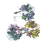
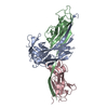
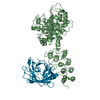
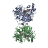
[English] 日本語
Yorodumi
- PDB-2os7: Caf1M periplasmic chaperone tetramer -

+
Open data


ID or keywords:

Loading...

-
Basic information

Entry
Database: PDB / ID: 2os7
TitleCaf1M periplasmic chaperone tetramer
ComponentsChaperone protein caf1M
KeywordsCHAPERONE / Immunoglobulin fold / Hetero beta barrel
Function / homology
Function and homology information


chaperone-mediated protein folding / cell wall organization / outer membrane-bounded periplasmic space
Similarity search - Function
Pili assembly chaperone, C-terminal / Pili assembly chaperone PapD, C-terminal domain / Pili assembly chaperone, bacterial / Pili assembly chaperone, conserved site / Pili assembly chaperone, C-terminal domain superfamily / Gram-negative pili assembly chaperone signature. / Pili assembly chaperone, N-terminal / Pili and flagellar-assembly chaperone, PapD N-terminal domain / PapD-like superfamily / Immunoglobulins ...Pili assembly chaperone, C-terminal / Pili assembly chaperone PapD, C-terminal domain / Pili assembly chaperone, bacterial / Pili assembly chaperone, conserved site / Pili assembly chaperone, C-terminal domain superfamily / Gram-negative pili assembly chaperone signature. / Pili assembly chaperone, N-terminal / Pili and flagellar-assembly chaperone, PapD N-terminal domain / PapD-like superfamily / Immunoglobulins / Immunoglobulin-like fold / Immunoglobulin-like / Sandwich / Mainly Beta
Similarity search - Domain/homology
Chaperone protein caf1M
Similarity search - Component
Biological speciesYersinia pestis (bacteria)
MethodX-RAY DIFFRACTION / SYNCHROTRON / MOLECULAR REPLACEMENT / Resolution: 2.9 Å
AuthorsKnight, S.D. / Zavialov, A.Z.
CitationJournal: MOL.MICROBIOL. / Year: 2007
Title: A novel self-capping mechanism controls aggregation of periplasmic chaperone Caf1M
Authors: Zavialov, A.Z. / Knight, S.D.
History
DepositionFeb 5, 2007Deposition site: RCSB / Processing site: RCSB
Revision 1.0Apr 17, 2007Provider: repository / Type: Initial release
Revision 1.1May 1, 2008Group: Version format compliance
Revision 1.2Jul 13, 2011Group: Advisory / Version format compliance
Revision 1.3Aug 30, 2023Group: Data collection / Database references / Refinement description
Category: chem_comp_atom / chem_comp_bond ...chem_comp_atom / chem_comp_bond / database_2 / pdbx_initial_refinement_model / struct_ncs_dom_lim
Item: _database_2.pdbx_DOI / _database_2.pdbx_database_accession ..._database_2.pdbx_DOI / _database_2.pdbx_database_accession / _struct_ncs_dom_lim.beg_auth_comp_id / _struct_ncs_dom_lim.end_auth_comp_id

-
Structure visualization

Structure viewerMolecule:
MolmilJmol/JSmol

Downloads & links

-
Assembly

Deposited unit
A: Chaperone protein caf1M
B: Chaperone protein caf1M
C: Chaperone protein caf1M
D: Chaperone protein caf1M
E: Chaperone protein caf1M
F: Chaperone protein caf1M


Theoretical massNumber of molelcules
Total (without water)157,9746
Polymers157,9746
Non-polymers00
Water00
1
A: Chaperone protein caf1M
B: Chaperone protein caf1M

A: Chaperone protein caf1M
B: Chaperone protein caf1M


Theoretical massNumber of molelcules
Total (without water)105,3164
Polymers105,3164
Non-polymers00
Water0
TypeNameSymmetry operationNumber
identity operation1_555x,y,z1
crystal symmetry operation2_555-x,-y,z1
2
C: Chaperone protein caf1M
D: Chaperone protein caf1M
E: Chaperone protein caf1M
F: Chaperone protein caf1M


Theoretical massNumber of molelcules
Total (without water)105,3164
Polymers105,3164
Non-polymers00
Water0
TypeNameSymmetry operationNumber
identity operation1_555x,y,z1
Buried area15480 Å2
ΔGint-84 kcal/mol
Surface area35230 Å2
MethodPISA
Unit cell
Length a, b, c (Å)141.092, 174.267, 60.580
Angle α, β, γ (deg.)90.00, 90.00, 90.00
Int Tables number18
Space group name H-MP21212
Noncrystallographic symmetry (NCS)NCS domain:
IDEns-IDDetails
11A
21C
31E
41A
51C
61E
71A
81C
91E
101A
111C
121E
131A
141C
151E
12B
22D
32F
42B
52D
62F
72B
82D
92F
102B
112D
122F
132B
142D
152F
162B
172D
182F
192B
202D
212F

NCS domain segments:

Refine code: 1

Dom-IDComponent-IDEns-IDBeg auth comp-IDBeg label comp-IDEnd auth comp-IDEnd label comp-IDAuth asym-IDLabel asym-IDAuth seq-IDLabel seq-ID
111GLYGLYLEULEUAA13 - 7813 - 78
211GLYGLYLEULEUCC13 - 7813 - 78
311GLYGLYLEULEUEE13 - 7813 - 78
421ILEILEPROPROAA80 - 8880 - 88
521ILEILEPROPROCC80 - 8880 - 88
621ILEILEPROPROEE80 - 8880 - 88
731ASPASPPROPROAA90 - 10390 - 103
831ASPASPPROPROCC90 - 10390 - 103
931ASPASPPROPROEE90 - 10390 - 103
1041VALVALPROPROAA126 - 205126 - 205
1141VALVALPROPROCC126 - 205126 - 205
1241VALVALPROPROEE126 - 205126 - 205
1351ASNASNASNASNAA213 - 232213 - 232
1451ASNASNASNASNCC213 - 232213 - 232
1551ASNASNASNASNEE213 - 232213 - 232
112ALAALAILEILEBB8 - 488 - 48
212ALAALAILEILEDD8 - 488 - 48
312ALAALAILEILEFF8 - 488 - 48
422PROPROALAALABB60 - 8160 - 81
522PROPROALAALADD60 - 8160 - 81
622PROPROALAALAFF60 - 8160 - 81
732LYSLYSPROPROBB91 - 10391 - 103
832LYSLYSPROPRODD91 - 10391 - 103
932LYSLYSPROPROFF91 - 10391 - 103
1042VALVALVALVALBB126 - 142126 - 142
1142VALVALVALVALDD126 - 142126 - 142
1242VALVALVALVALFF126 - 142126 - 142
1352PHEPHETRPTRPBB154 - 160154 - 160
1452PHEPHETRPTRPDD154 - 160154 - 160
1552PHEPHETRPTRPFF154 - 160154 - 160
1662TYRTYRLEULEUBB176 - 204176 - 204
1762TYRTYRLEULEUDD176 - 204176 - 204
1862TYRTYRLEULEUFF176 - 204176 - 204
1972ASNASNASNASNBB213 - 232213 - 232
2072ASNASNASNASNDD213 - 232213 - 232
2172ASNASNASNASNFF213 - 232213 - 232

NCS ensembles :
ID
1
2
DetailsThe biological assembly is a tetramer of Caf1M chaperone protomers. The assymetric unit contains one-and-a-half tetramer.

-
Components

#1: Protein
Chaperone protein caf1M / Capsule protein fraction 1


Mass: 26329.012 Da / Num. of mol.: 6
Source method: isolated from a genetically manipulated source
Source: (gene. exp.) Yersinia pestis (bacteria) / Gene: caf1M / Plasmid: pTCA1 / Production host: Escherichia coli (E. coli) / Strain (production host): B834 (DE3) / References: UniProt: P26926

-
Experimental details

-
Experiment

ExperimentMethod: X-RAY DIFFRACTION / Number of used crystals: 1

-
Sample preparation

CrystalDensity Matthews: 2.35 Å3/Da / Density % sol: 47.75 %
Crystal growTemperature: 293 K / Method: vapor diffusion, hanging drop / pH: 8
Details: 8% PEG 6000 in 50 mM Tris-HCl pH 8.0-8.3, VAPOR DIFFUSION, HANGING DROP, temperature 293K

-
Data collection

DiffractionMean temperature: 100 K
Diffraction sourceSource: SYNCHROTRON / Site: ESRF / Beamline: ID14-2 / Wavelength: 0.933 Å
DetectorType: MARMOSAIC 225 mm CCD / Detector: CCD / Date: Jan 1, 2004
RadiationProtocol: SINGLE WAVELENGTH / Monochromatic (M) / Laue (L): M / Scattering type: x-ray
Radiation wavelengthWavelength: 0.933 Å / Relative weight: 1
ReflectionResolution: 2.9→200 Å / Num. obs: 33968 / % possible obs: 99.9 % / Redundancy: 6 % / Biso Wilson estimate: 65.1 Å2 / Rmerge(I) obs: 0.165 / Net I/σ(I): 3.8
Reflection shellResolution: 2.9→3.16 Å / Redundancy: 6 % / Rmerge(I) obs: 0.4 / Mean I/σ(I) obs: 3.8 / Num. unique all: 3722 / % possible all: 99.9

-
Processing

Software
NameVersionClassification
REFMAC5.2.0005refinement
HKL-2000data collection
MOSFLMdata reduction
CCP4(SCALA)data scaling
MOLREPphasing
RefinementMethod to determine structure: MOLECULAR REPLACEMENT
Starting model: Caf1M (chain A) from pdb entry 1p5v
Resolution: 2.9→30 Å / Cor.coef. Fo:Fc: 0.892 / Cor.coef. Fo:Fc free: 0.837 / SU B: 44.81 / SU ML: 0.387 / TLS residual ADP flag: LIKELY RESIDUAL / Isotropic thermal model: Isotropic / Cross valid method: THROUGHOUT / σ(F): 0 / σ(I): 0 / ESU R Free: 0.477 / Stereochemistry target values: MAXIMUM LIKELIHOOD
Details: TLS refinment using two TLS groups (tetramer in au + half-tetramer in au) Tight NCS restraints (two NCS groups: chains A,C,E and chains B,D,F)
RfactorNum. reflection% reflectionSelection details
Rfree0.29843 1720 5.1 %RANDOM
Rwork0.24906 ---
obs0.25157 32199 99.83 %-
all-32199 --
Solvent computationIon probe radii: 0.8 Å / Shrinkage radii: 0.8 Å / VDW probe radii: 1.2 Å / Solvent model: BABINET MODEL WITH MASK
Displacement parametersBiso mean: 45.389 Å2
Baniso -1Baniso -2Baniso -3
1--5.28 Å20 Å20 Å2
2--1.45 Å20 Å2
3---3.82 Å2
Refine analyzeLuzzati coordinate error free: 0.477 Å
Refinement stepCycle: LAST / Resolution: 2.9→30 Å
ProteinNucleic acidLigandSolventTotal
Num. atoms9426 0 0 0 9426
Refine LS restraints
Refine-IDTypeDev idealDev ideal targetNumber
X-RAY DIFFRACTIONr_bond_refined_d0.0170.0229662
X-RAY DIFFRACTIONr_bond_other_d
X-RAY DIFFRACTIONr_angle_refined_deg1.6541.96713085
X-RAY DIFFRACTIONr_angle_other_deg
X-RAY DIFFRACTIONr_dihedral_angle_1_deg7.74751172
X-RAY DIFFRACTIONr_dihedral_angle_2_deg36.57624.532417
X-RAY DIFFRACTIONr_dihedral_angle_3_deg20.486151650
X-RAY DIFFRACTIONr_dihedral_angle_4_deg20.6471547
X-RAY DIFFRACTIONr_chiral_restr0.0980.21421
X-RAY DIFFRACTIONr_gen_planes_refined0.0050.027261
X-RAY DIFFRACTIONr_gen_planes_other
X-RAY DIFFRACTIONr_nbd_refined0.2380.23732
X-RAY DIFFRACTIONr_nbd_other
X-RAY DIFFRACTIONr_nbtor_refined0.3220.26353
X-RAY DIFFRACTIONr_nbtor_other
X-RAY DIFFRACTIONr_xyhbond_nbd_refined0.150.2297
X-RAY DIFFRACTIONr_xyhbond_nbd_other
X-RAY DIFFRACTIONr_metal_ion_refined
X-RAY DIFFRACTIONr_metal_ion_other
X-RAY DIFFRACTIONr_symmetry_vdw_refined0.2910.289
X-RAY DIFFRACTIONr_symmetry_vdw_other
X-RAY DIFFRACTIONr_symmetry_hbond_refined0.2460.210
X-RAY DIFFRACTIONr_symmetry_hbond_other
X-RAY DIFFRACTIONr_symmetry_metal_ion_refined
X-RAY DIFFRACTIONr_symmetry_metal_ion_other
X-RAY DIFFRACTIONr_mcbond_it0.5411.56083
X-RAY DIFFRACTIONr_mcbond_other
X-RAY DIFFRACTIONr_mcangle_it0.94429639
X-RAY DIFFRACTIONr_scbond_it1.25734110
X-RAY DIFFRACTIONr_scangle_it2.0914.53446
X-RAY DIFFRACTIONr_rigid_bond_restr
X-RAY DIFFRACTIONr_sphericity_free
X-RAY DIFFRACTIONr_sphericity_bonded
Refine LS restraints NCS

Refine-ID: X-RAY DIFFRACTION

Ens-IDDom-IDAuth asym-IDNumberTypeRms dev position (Å)Weight position
11A1486tight positional0.070.05
12C1486tight positional0.060.05
13E1486tight positional0.060.05
21B1164tight positional0.060.05
22D1164tight positional0.060.05
23F1164tight positional0.060.05
11A1486tight thermal0.110.5
12C1486tight thermal0.110.5
13E1486tight thermal0.110.5
21B1164tight thermal0.10.5
22D1164tight thermal0.10.5
23F1164tight thermal0.10.5
LS refinement shellResolution: 2.9→3.057 Å / Total num. of bins used: 10
RfactorNum. reflection% reflection
Rfree0.356 247 -
Rwork0.321 4605 -
obs--99.88 %
Refinement TLS params.

Method: refined / Refine-ID: X-RAY DIFFRACTION

IDL112)L122)L132)L222)L232)L332)S11 (Å °)S12 (Å °)S13 (Å °)S21 (Å °)S22 (Å °)S23 (Å °)S31 (Å °)S32 (Å °)S33 (Å °)T112)T122)T132)T222)T232)T332)Origin x (Å)Origin y (Å)Origin z (Å)
11.2353-0.23490.0350.8306-0.06170.7275-0.01570.08870.2381-0.08310.0131-0.0792-0.12830.05290.0026-0.0692-0.0250.0207-0.16260.0289-0.06260.751524.064617.184
20.59560.0854-0.16871.284-0.1190.1149-0.0081-0.0794-0.05710.0848-0.005-0.1050.0050.06110.0131-0.0405-0.0101-0.02230.00780.0146-0.118946.68010.694724.286
Refinement TLS group
IDRefine-IDRefine TLS-IDAuth asym-IDLabel asym-IDAuth seq-IDLabel seq-ID
1X-RAY DIFFRACTION1AA3 - 2333 - 233
2X-RAY DIFFRACTION1BB6 - 2336 - 233
3X-RAY DIFFRACTION2CC3 - 2333 - 233
4X-RAY DIFFRACTION2DD6 - 2326 - 232
5X-RAY DIFFRACTION2EE3 - 2323 - 232
6X-RAY DIFFRACTION2FF6 - 2336 - 233

+
About Yorodumi

-
News

-
Feb 9, 2022. New format data for meta-information of EMDB entries

New format data for meta-information of EMDB entries

  • Version 3 of the EMDB header file is now the official format.
  • The previous official version 1.9 will be removed from the archive.

Related info.:EMDB header

External links:wwPDB to switch to version 3 of the EMDB data model

-
Aug 12, 2020. Covid-19 info

Covid-19 info

URL: https://pdbjlvh1.pdbj.org/emnavi/covid19.php

New page: Covid-19 featured information page in EM Navigator.

Related info.:Covid-19 info / Mar 5, 2020. Novel coronavirus structure data

+
Mar 5, 2020. Novel coronavirus structure data

Novel coronavirus structure data

Related info.:Yorodumi Speices / Aug 12, 2020. Covid-19 info

External links:COVID-19 featured content - PDBj / Molecule of the Month (242):Coronavirus Proteases

+
Jan 31, 2019. EMDB accession codes are about to change! (news from PDBe EMDB page)

EMDB accession codes are about to change! (news from PDBe EMDB page)

  • The allocation of 4 digits for EMDB accession codes will soon come to an end. Whilst these codes will remain in use, new EMDB accession codes will include an additional digit and will expand incrementally as the available range of codes is exhausted. The current 4-digit format prefixed with “EMD-” (i.e. EMD-XXXX) will advance to a 5-digit format (i.e. EMD-XXXXX), and so on. It is currently estimated that the 4-digit codes will be depleted around Spring 2019, at which point the 5-digit format will come into force.
  • The EM Navigator/Yorodumi systems omit the EMD- prefix.

Related info.:Q: What is EMD? / ID/Accession-code notation in Yorodumi/EM Navigator

External links:EMDB Accession Codes are Changing Soon! / Contact to PDBj

+
Jul 12, 2017. Major update of PDB

Major update of PDB

  • wwPDB released updated PDB data conforming to the new PDBx/mmCIF dictionary.
  • This is a major update changing the version number from 4 to 5, and with Remediation, in which all the entries are updated.
  • In this update, many items about electron microscopy experimental information are reorganized (e.g. em_software).
  • Now, EM Navigator and Yorodumi are based on the updated data.

External links:wwPDB Remediation / Enriched Model Files Conforming to OneDep Data Standards Now Available in the PDB FTP Archive

-
Yorodumi

Thousand views of thousand structures

  • Yorodumi is a browser for structure data from EMDB, PDB, SASBDB, etc.
  • This page is also the successor to EM Navigator detail page, and also detail information page/front-end page for Omokage search.
  • The word "yorodu" (or yorozu) is an old Japanese word meaning "ten thousand". "mi" (miru) is to see.

Related info.:EMDB / PDB / SASBDB / Comparison of 3 databanks / Yorodumi Search / Aug 31, 2016. New EM Navigator & Yorodumi / Yorodumi Papers / Jmol/JSmol / Function and homology information / Changes in new EM Navigator and Yorodumi

Read more