 Movie
Movie Controller


+ Open data

- Basic information
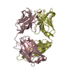
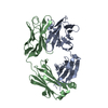
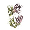
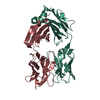
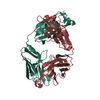
| Entry | Database: PDB / ID: 2orb | ||||||
|---|---|---|---|---|---|---|---|
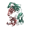
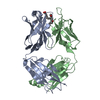
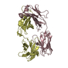
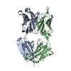
| Title | The structure of the anti-c-myc antibody 9E10 Fab fragment | ||||||
 Components Components | (Monoclonal anti-c-myc antibody 9E10) x 2 | ||||||
 Keywords Keywords | IMMUNE SYSTEM / ANTIGEN-ANTIBODY COMPLEX / ANTIGEN RECOGNITION / long CDR H3 | ||||||
| Function / homology | Immunoglobulins / Immunoglobulin-like / Sandwich / Mainly Beta Function and homology information Function and homology information | ||||||
| Biological species |  | ||||||
| Method |  X-RAY DIFFRACTION / SYNCHROTRON / MOLECULAR REPLACEMENT / Resolution: 2.2 Å X-RAY DIFFRACTION / SYNCHROTRON / MOLECULAR REPLACEMENT / Resolution: 2.2 Å | ||||||
 Authors Authors | Krauss, N. / Scheerer, P. / Hoehne, W. | ||||||
 Citation Citation | Journal: Proteins / Year: 2008 Title: The structure of the anti-c-myc antibody 9E10 Fab fragment/epitope peptide complex reveals a novel binding mode dominated by the heavy chain hypervariable loops. Authors: Krauss, N. / Wessner, H. / Welfle, K. / Welfle, H. / Scholz, C. / Seifert, M. / Zubow, K. / Ay, J. / Hahn, M. / Scheerer, P. / Skerra, A. / Hohne, W. | ||||||
| History |
|
- Structure visualization
| Structure viewer | Molecule: MolmilJmol/JSmol |
|---|
- Downloads & links
-Download
| PDBx/mmCIF format | 2orb.cif.gz | 177.3 KB | Display | PDBx/mmCIF format |
|---|---|---|---|---|
| PDB format | pdb2orb.ent.gz | 140.5 KB | Display | PDB format |
| PDBx/mmJSON format | 2orb.json.gz | Tree view | PDBx/mmJSON format | |
| Others |  Other downloads Other downloads |
-Validation report
| Arichive directory | https://data.pdbj.org/pub/pdb/validation_reports/or/2orbftp://data.pdbj.org/pub/pdb/validation_reports/or/2orb | HTTPS FTP |
|---|
-Related structure data
| Related structure data |  2or9SC S: Starting model for refinement C: citing same article ( |
|---|---|
| Similar structure data |


-Links
 PDBj
PDBj

- Assembly
| Deposited unit | 
| ||||||||
|---|---|---|---|---|---|---|---|---|---|
| 1 | 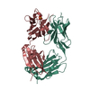
| ||||||||
| 2 | 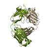
| ||||||||
| Unit cell |
|
-Components
| #1: Antibody | Mass: 24001.543 Da / Num. of mol.: 2 / Fragment: light chain of antigen binding fragment, Fab / Source method: isolated from a natural source / Source: (natural) #2: Antibody | Mass: 24899.799 Da / Num. of mol.: 2 / Fragment: heavy chain of antigen binding fragment, Fab / Source method: isolated from a natural source / Source: (natural) #3: Chemical |   Mass: 96.063 Da / Num. of mol.: 2 / Source method: obtained synthetically / Formula: SO4 Mass: 96.063 Da / Num. of mol.: 2 / Source method: obtained synthetically / Formula: SO4#4: Water | ChemComp-HOH / |  Mass: 18.015 Da / Num. of mol.: 257 / Source method: isolated from a natural source / Formula: H2O Mass: 18.015 Da / Num. of mol.: 257 / Source method: isolated from a natural source / Formula: H2OHas protein modification | Y | |
|---|
-Experimental details
-Experiment
| Experiment | Method: X-RAY DIFFRACTION / Number of used crystals: 1 |
|---|
- Sample preparation
| Crystal | Density Matthews: 2.09 Å3/Da / Density % sol: 41.26 % |
|---|---|
| Crystal grow | Temperature: 291 K / Method: vapor diffusion, hanging drop / pH: 3.05 Details: 15% PEG 3350, 0.2M ammonium sulfate, 0.1 M sodium citrate buffer, pH 3.05, 0.02% sodium azide, VAPOR DIFFUSION, HANGING DROP, temperature 291K |
-Data collection
| Diffraction | Mean temperature: 100 K |
|---|---|
| Diffraction source | Source: SYNCHROTRON / Site: EMBL/DESY, HAMBURG  / Beamline: X11 / Wavelength: 0.811 / Beamline: X11 / Wavelength: 0.811 |
| Detector | Type: MAR CCD 165 mm / Detector: CCD / Date: Feb 25, 2004 |
| Radiation | Monochromator: TRIANGULAR MONOCHROMATOR / Protocol: SINGLE WAVELENGTH / Monochromatic (M) / Laue (L): M / Scattering type: x-ray |
| Radiation wavelength | Wavelength: 0.811 Å / Relative weight: 1 |
| Reflection | Resolution: 2.2→25 Å / Num. all: 37773 / Num. obs: 37773 / % possible obs: 91.5 % / Observed criterion σ(F): 0 / Observed criterion σ(I): -3 / Redundancy: 3 % / Biso Wilson estimate: 41.2 Å2 / Rsym value: 0.046 / Net I/σ(I): 19.9 |
| Reflection shell | Resolution: 2.2→2.24 Å / Redundancy: 2.5 % / Mean I/σ(I) obs: 2.7 / Num. unique all: 1278 / Rsym value: 0.345 / % possible all: 62.1 |
- Processing
| Software |
| |||||||||||||||||||||||||
|---|---|---|---|---|---|---|---|---|---|---|---|---|---|---|---|---|---|---|---|---|---|---|---|---|---|---|
| Refinement | Method to determine structure: MOLECULAR REPLACEMENT Starting model: PDB entry 2OR9 Resolution: 2.2→25 Å / Isotropic thermal model: Isotropic / Cross valid method: THROUGHOUT / σ(F): 0 / Stereochemistry target values: Engh & Huber
| |||||||||||||||||||||||||
| Displacement parameters | Biso mean: 46.8 Å2
| |||||||||||||||||||||||||
| Refine analyze |
| |||||||||||||||||||||||||
| Refinement step | Cycle: LAST / Resolution: 2.2→25 Å
| |||||||||||||||||||||||||
| Refine LS restraints |
| |||||||||||||||||||||||||
| LS refinement shell | Resolution: 2.2→2.28 Å
|
