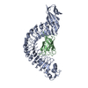
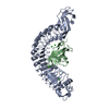
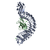
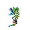
Entry Database : PDB / ID : 2omxTitle Crystal structure of InlA S192N G194S+S/hEC1 complex Epithelial-cadherin Internalin-A Keywords / / / / / Function / homology Function Domain/homology Component
/ / / / / / / / / / / / / / / / / / / / / / / / / / / / / / / / / / / / / / / / / / / / / / / / / / / / / / / / / / / / / / / / / / / / / / / / / / / / / / / / / / / / / / / / / / / / / / / / / / / / / / / / / / / / / / / / / / / / / / / / / / / / / / / / / / / / / / / / / / / / Biological species Listeria monocytogenes (bacteria)Homo sapiens (human)Method / / / Resolution : 1.7 Å Authors Wollert, T. / Heinz, D.W. / Schubert, W.D. Journal : Proc.Natl.Acad.Sci.Usa / Year : 2007Title : Thermodynamically reengineering the listerial invasion complex InlA/E-cadherin.Authors : Wollert, T. / Heinz, D.W. / Schubert, W.D. History Deposition Jan 23, 2007 Deposition site / Processing site Revision 1.0 Aug 28, 2007 Provider / Type Revision 1.1 Jul 13, 2011 Group / Source and taxonomy / Version format complianceRevision 1.2 Oct 18, 2017 Group / Refinement description / Category / softwareRevision 1.3 Oct 20, 2021 Group Advisory / Data collection ... Advisory / Data collection / Database references / Derived calculations Category database_2 / diffrn_source ... database_2 / diffrn_source / pdbx_unobs_or_zero_occ_atoms / struct_ref_seq_dif / struct_site Item _database_2.pdbx_DOI / _database_2.pdbx_database_accession ... _database_2.pdbx_DOI / _database_2.pdbx_database_accession / _diffrn_source.pdbx_synchrotron_site / _struct_ref_seq_dif.details / _struct_site.pdbx_auth_asym_id / _struct_site.pdbx_auth_comp_id / _struct_site.pdbx_auth_seq_id Revision 1.4 Aug 30, 2023 Group / Refinement descriptionCategory / chem_comp_bond / pdbx_initial_refinement_model
Show all Show less
 Movie
Movie Controller


 Open data
 Basic information
 Components
 Keywords
 Function and homology information
 Listeria monocytogenes (bacteria)
 Homo sapiens (human)
 X-RAY DIFFRACTION /
  Authors
 Citation
 Structure visualization
 Downloads & links
 Other downloads


 PDBj
PDBj


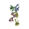
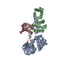
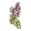
 Assembly
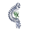

 Mass: 35.453 Da / Num. of mol.: 1 / Source method: obtained synthetically / Formula: Cl
 Mass: 40.078 Da / Num. of mol.: 1 / Source method: obtained synthetically / Formula: Ca
 Mass: 18.015 Da / Num. of mol.: 707 / Source method: isolated from a natural source / Formula: H2O
 Sample preparation
 / Beamline: X11 / Wavelength: 0.81 Å
 Processing