 Movie
Movie Controller

[English] 日本語
 Yorodumi
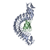
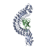
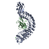
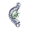
- PDB-1o6s: Internalin (Listeria monocytogenes) / E-Cadherin (human) Recognit... -
+ Open data

- Basic information
| Entry | Database: PDB / ID: 1o6s | ||||||
|---|---|---|---|---|---|---|---|
| Title | Internalin (Listeria monocytogenes) / E-Cadherin (human) Recognition Complex | ||||||
 Components Components |
| ||||||
 Keywords Keywords | BACTERIAL INFECTION / LEUCINE RICH REPEAT / CELL ADHESION / CELL-WALL SURFACE PROTEIN | ||||||
| Function / homology |  Function and homology information Function and homology informationresponse to heparin / desmosome assembly / response to Gram-positive bacterium / pituitary gland development / Regulation of MITF-M-dependent genes involved in extracellular matrix, focal adhesion and epithelial-to-mesenchymal transition / gamma-catenin binding / desmosome / cell adhesion mediator activity / Regulation of CDH1 mRNA translation by microRNAs / negative regulation of axon extension ...response to heparin / desmosome assembly / response to Gram-positive bacterium / pituitary gland development / Regulation of MITF-M-dependent genes involved in extracellular matrix, focal adhesion and epithelial-to-mesenchymal transition / gamma-catenin binding / desmosome / cell adhesion mediator activity / Regulation of CDH1 mRNA translation by microRNAs / negative regulation of axon extension / cellular response to indole-3-methanol / calcium-dependent cell-cell adhesion / regulation of protein catabolic process at postsynapse, modulating synaptic transmission / flotillin complex / Developmental Lineage of Mammary Stem Cells / cell-cell adhesion mediated by cadherin / Formation of definitive endoderm / adherens junction organization / Apoptotic cleavage of cell adhesion proteins / catenin complex / Regulation of CDH1 Function / cell-cell junction assembly / Adherens junctions interactions / GTPase activating protein binding / negative regulation of cell-cell adhesion / ankyrin binding / cellular response to lithium ion / Regulation of CDH1 posttranslational processing and trafficking to plasma membrane / apical junction complex / homophilic cell-cell adhesion / Developmental Lineage of Mammary Gland Myoepithelial Cells / lateral plasma membrane / Integrin cell surface interactions / RHO GTPases activate IQGAPs / Degradation of the extracellular matrix / positive regulation of protein localization / Transcriptional and post-translational regulation of MITF-M expression and activity / synapse assembly / cell adhesion molecule binding / peptidoglycan-based cell wall / protein tyrosine kinase binding / negative regulation of cell migration / InlA-mediated entry of Listeria monocytogenes into host cells / protein localization to plasma membrane / adherens junction / trans-Golgi network / cell-cell adhesion / positive regulation of protein import into nucleus / beta-catenin binding / Golgi lumen / Degradation of CDH1 / cell morphogenesis / response to toxic substance / cytoplasmic side of plasma membrane / Immunoregulatory interactions between a Lymphoid and a non-Lymphoid cell / neuron projection development / cell junction / cell migration / lamellipodium / actin cytoskeleton / regulation of gene expression / early endosome membrane / postsynapse / cadherin binding / response to xenobiotic stimulus / endoplasmic reticulum lumen / Golgi membrane / lysosomal membrane / calcium ion binding / endoplasmic reticulum membrane / positive regulation of DNA-templated transcription / perinuclear region of cytoplasm / glutamatergic synapse / extracellular exosome / extracellular region / membrane / identical protein binding / plasma membrane / cytoplasm Similarity search - Function | ||||||
| Biological species |  LISTERIA MONOCYTOGENES (bacteria) LISTERIA MONOCYTOGENES (bacteria) HOMO SAPIENS (human) HOMO SAPIENS (human) | ||||||
| Method |  X-RAY DIFFRACTION / SYNCHROTRON / MOLECULAR REPLACEMENT / Resolution: 1.8 Å X-RAY DIFFRACTION / SYNCHROTRON / MOLECULAR REPLACEMENT / Resolution: 1.8 Å | ||||||
 Authors Authors | Schubert, W.-D. / Urbanke, C. / Ziehm, T. / Beier, V. / Machner, M.P. / Domann, E. / Wehland, J. / Chakraborty, T. / Heinz, D.W. | ||||||
 Citation Citation | Journal: Cell(Cambridge,Mass.) / Year: 2002 Title: Structure of Internalin, a Major Invasion Protein of Listeria Monocytogenes, in Complex with its Human Receptor E-Cadherin Authors: Schubert, W.-D. / Urbanke, C. / Ziehm, T. / Beier, V. / Machner, M.P. / Domann, E. / Wehland, J. / Chakraborty, T. / Heinz, D.W. | ||||||
| History |
|
- Structure visualization
| Structure viewer | Molecule: MolmilJmol/JSmol |
|---|
- Downloads & links
-Download
| PDBx/mmCIF format | 1o6s.cif.gz | 139.2 KB | Display | PDBx/mmCIF format |
|---|---|---|---|---|
| PDB format | pdb1o6s.ent.gz | 108.2 KB | Display | PDB format |
| PDBx/mmJSON format | 1o6s.json.gz | Tree view | PDBx/mmJSON format | |
| Others |  Other downloads Other downloads |
-Validation report
| Arichive directory | https://data.pdbj.org/pub/pdb/validation_reports/o6/1o6sftp://data.pdbj.org/pub/pdb/validation_reports/o6/1o6s | HTTPS FTP |
|---|
-Related structure data


-Links
 PDBj
PDBj


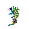
- Assembly
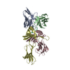
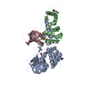
| Deposited unit | 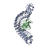
| ||||||||
|---|---|---|---|---|---|---|---|---|---|
| 1 |
| ||||||||
| Unit cell |
|
-Components
| #1: Protein | Mass: 50239.133 Da / Num. of mol.: 1 / Fragment: FUNCTIONAL DOMAIN, RESIDUES 36-496 Source method: isolated from a genetically manipulated source Source: (gene. exp.) LISTERIA MONOCYTOGENES (bacteria) / Strain: EGD-E / Variant: SEROVAR 1/2A / Plasmid: PGEX-6P-1 / Production host: | ||||||
|---|---|---|---|---|---|---|---|
| #2: Protein | Mass: 11523.009 Da / Num. of mol.: 1 / Fragment: N-TERMINAL DOMAIN, RESIDUES 156-253 Source method: isolated from a genetically manipulated source Source: (gene. exp.) HOMO SAPIENS (human) / Plasmid: PGEX-6P-1 / Production host: | ||||||
| #3: Chemical |   Mass: 40.078 Da / Num. of mol.: 2 / Source method: obtained synthetically / Formula: Ca Mass: 40.078 Da / Num. of mol.: 2 / Source method: obtained synthetically / Formula: Ca#4: Chemical |   Mass: 35.453 Da / Num. of mol.: 3 / Source method: obtained synthetically / Formula: Cl Mass: 35.453 Da / Num. of mol.: 3 / Source method: obtained synthetically / Formula: Cl#5: Water | ChemComp-HOH / |  Mass: 18.015 Da / Num. of mol.: 537 / Source method: isolated from a natural source / Formula: H2O Mass: 18.015 Da / Num. of mol.: 537 / Source method: isolated from a natural source / Formula: H2OSequence details | FIRST FIVE RESIDUES (GPLGS) INTRODUCED | |
-Experimental details
-Experiment
| Experiment | Method: X-RAY DIFFRACTION / Number of used crystals: 1 |
|---|
- Sample preparation
| Crystal | Density Matthews: 2.14 Å3/Da / Density % sol: 42 % | ||||||||||||||||||||||||||||||||||||||||||
|---|---|---|---|---|---|---|---|---|---|---|---|---|---|---|---|---|---|---|---|---|---|---|---|---|---|---|---|---|---|---|---|---|---|---|---|---|---|---|---|---|---|---|---|
| Crystal grow | Method: vapor diffusion, hanging drop / pH: 6 Details: VAPOUR DIFFUSION / HANGING DROP 10 MG/ML IN 10 MM HEPES PH 7.0, 26% PEG 4000, 100 MM MES/TRIS PH 7.0, 100 MM SODIUM ACETATE, 50 MM CACL2 | ||||||||||||||||||||||||||||||||||||||||||
| Crystal grow | *PLUS Temperature: 20 ℃ / pH: 7 / Method: vapor diffusion, hanging drop | ||||||||||||||||||||||||||||||||||||||||||
| Components of the solutions | *PLUS
|
-Data collection
| Diffraction | Mean temperature: 100 K |
|---|---|
| Diffraction source | Source: SYNCHROTRON / Site: MPG/DESY, HAMBURG  / Beamline: BW6 / Wavelength: 1.05 / Beamline: BW6 / Wavelength: 1.05 |
| Detector | Type: MARRESEARCH / Detector: CCD / Date: Aug 15, 2002 |
| Radiation | Protocol: SINGLE WAVELENGTH / Monochromatic (M) / Laue (L): M / Scattering type: x-ray |
| Radiation wavelength | Wavelength: 1.05 Å / Relative weight: 1 |
| Reflection | Resolution: 1.8→25.1 Å / Num. obs: 50336 / % possible obs: 92 % / Redundancy: 6.2 % / Rmerge(I) obs: 0.062 / Net I/σ(I): 14.4 |
| Reflection shell | Resolution: 1.8→1.86 Å / Redundancy: 4.5 % / Rmerge(I) obs: 0.385 / Mean I/σ(I) obs: 3.4 / % possible all: 84.3 |
| Reflection | *PLUS Highest resolution: 1.8 Å / Lowest resolution: 25 Å / Num. obs: 46306 |
| Reflection shell | *PLUS % possible obs: 84.3 % |
- Processing
| Software |
| ||||||||||||||||||||||||||||||||||||||||||||||||||||||||||||||||||||||||||||||||||||||||||||||||||||||||||||||||||||||||||||||||||||||||||||||||||||||||||||||||||||||||||||||||||||||
|---|---|---|---|---|---|---|---|---|---|---|---|---|---|---|---|---|---|---|---|---|---|---|---|---|---|---|---|---|---|---|---|---|---|---|---|---|---|---|---|---|---|---|---|---|---|---|---|---|---|---|---|---|---|---|---|---|---|---|---|---|---|---|---|---|---|---|---|---|---|---|---|---|---|---|---|---|---|---|---|---|---|---|---|---|---|---|---|---|---|---|---|---|---|---|---|---|---|---|---|---|---|---|---|---|---|---|---|---|---|---|---|---|---|---|---|---|---|---|---|---|---|---|---|---|---|---|---|---|---|---|---|---|---|---|---|---|---|---|---|---|---|---|---|---|---|---|---|---|---|---|---|---|---|---|---|---|---|---|---|---|---|---|---|---|---|---|---|---|---|---|---|---|---|---|---|---|---|---|---|---|---|---|---|
| Refinement | Method to determine structure: MOLECULAR REPLACEMENT / Resolution: 1.8→69.01 Å / Cor.coef. Fo:Fc: 0.967 / Cor.coef. Fo:Fc free: 0.946 / SU B: 3.368 / SU ML: 0.102 / Cross valid method: THROUGHOUT / ESU R: 0.148 / ESU R Free: 0.142 / Stereochemistry target values: MAXIMUM LIKELIHOOD / Details: HYDROGENS HAVE BEEN ADDED IN THE RIDING POSITIONS
| ||||||||||||||||||||||||||||||||||||||||||||||||||||||||||||||||||||||||||||||||||||||||||||||||||||||||||||||||||||||||||||||||||||||||||||||||||||||||||||||||||||||||||||||||||||||
| Solvent computation | Ion probe radii: 0.8 Å / Shrinkage radii: 0.8 Å / VDW probe radii: 1.4 Å / Solvent model: BABINET MODEL WITH MASK | ||||||||||||||||||||||||||||||||||||||||||||||||||||||||||||||||||||||||||||||||||||||||||||||||||||||||||||||||||||||||||||||||||||||||||||||||||||||||||||||||||||||||||||||||||||||
| Displacement parameters | Biso mean: 23.05 Å2
| ||||||||||||||||||||||||||||||||||||||||||||||||||||||||||||||||||||||||||||||||||||||||||||||||||||||||||||||||||||||||||||||||||||||||||||||||||||||||||||||||||||||||||||||||||||||
| Refinement step | Cycle: LAST / Resolution: 1.8→69.01 Å
| ||||||||||||||||||||||||||||||||||||||||||||||||||||||||||||||||||||||||||||||||||||||||||||||||||||||||||||||||||||||||||||||||||||||||||||||||||||||||||||||||||||||||||||||||||||||
| Refine LS restraints |
|