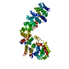
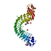
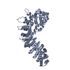
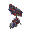
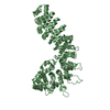
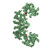
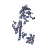
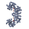
[English] 日本語
Yorodumi
- PDB-1o6v: Internalin (INLA, Listeria monocytogenes) - functional domain, un... -

+
Open data


ID or keywords:

Loading...

-
Basic information

Entry
Database: PDB / ID: 1o6v
TitleInternalin (INLA, Listeria monocytogenes) - functional domain, uncomplexed
ComponentsINTERNALIN A
KeywordsCELL ADHESION / BACTERIAL INFECTION / EXTRACELLULAR RECOGNITION / CELL WALL ATTACHED / LEUCINE RICH REPEAT
Function / homology
Function and homology information


InlA-mediated entry of Listeria monocytogenes into host cells / peptidoglycan-based cell wall / extracellular region
Similarity search - Function
: / Listeria/Bacterioides repeat / Listeria-Bacteroides repeat domain superfamily / Listeria-Bacteroides repeat domain (List_Bact_rpt) / Leucine-rich repeat-containing adjacent domain / LRR adjacent / Internalin, N-terminal / Bacterial adhesion/invasion protein N terminal / Immunoglobulin-like - #1220 / Copper resistance protein CopC/internalin, immunoglobulin-like ...: / Listeria/Bacterioides repeat / Listeria-Bacteroides repeat domain superfamily / Listeria-Bacteroides repeat domain (List_Bact_rpt) / Leucine-rich repeat-containing adjacent domain / LRR adjacent / Internalin, N-terminal / Bacterial adhesion/invasion protein N terminal / Immunoglobulin-like - #1220 / Copper resistance protein CopC/internalin, immunoglobulin-like / Leucine rich repeat 4 / Leucine Rich repeats (2 copies) / Leucine-rich repeat, LRR (right-handed beta-alpha superhelix) / Ribonuclease Inhibitor / Leucine-rich repeat, SDS22-like subfamily / Alpha-Beta Horseshoe / LPXTG cell wall anchor motif / Gram-positive cocci surface proteins LPxTG motif profile. / LPXTG cell wall anchor domain / Leucine-rich repeat, typical subtype / Leucine-rich repeats, typical (most populated) subfamily / Leucine-rich repeat profile. / Leucine-rich repeat / Leucine-rich repeat domain superfamily / Immunoglobulin E-set / Immunoglobulin-like / Sandwich / Mainly Beta / Alpha Beta
Similarity search - Domain/homology
Internalin A / Internalin A
Similarity search - Component
Biological speciesLISTERIA MONOCYTOGENES (bacteria)
MethodX-RAY DIFFRACTION / SYNCHROTRON / MOLECULAR REPLACEMENT / Resolution: 1.5 Å
AuthorsSchubert, W.-D. / Urbanke, C. / Ziehm, T. / Beier, V. / Machner, M.P. / Domann, E. / Wehland, J. / Chakraborty, T. / Heinz, D.W.
CitationJournal: Cell(Cambridge,Mass.) / Year: 2002
Title: Structure of Internalin, a Major Invasion Protein of Listeria Monocytogenes, in Complex with its Human Receptor E-Cadherin
Authors: Schubert, W.-D. / Urbanke, C. / Ziehm, T. / Beier, V. / Machner, M.P. / Domann, E. / Wehland, J. / Chakraborty, T. / Heinz, D.W.
History
DepositionOct 16, 2002Deposition site: PDBE / Processing site: PDBE
Revision 1.0Dec 23, 2002Provider: repository / Type: Initial release
Revision 1.1Aug 1, 2012Group: Advisory / Atomic model ...Advisory / Atomic model / Database references / Derived calculations / Non-polymer description / Other / Refinement description / Structure summary / Version format compliance
Revision 1.2May 8, 2019Group: Advisory / Data collection ...Advisory / Data collection / Experimental preparation / Other
Category: database_PDB_rev / database_PDB_rev_record ...database_PDB_rev / database_PDB_rev_record / exptl_crystal_grow / pdbx_database_proc / pdbx_database_status / pdbx_validate_polymer_linkage
Item: _exptl_crystal_grow.method / _pdbx_database_status.recvd_author_approval ..._exptl_crystal_grow.method / _pdbx_database_status.recvd_author_approval / _pdbx_validate_polymer_linkage.dist / _pdbx_validate_polymer_linkage.label_alt_id_1 / _pdbx_validate_polymer_linkage.label_alt_id_2
Revision 1.3May 8, 2024Group: Data collection / Database references ...Data collection / Database references / Derived calculations / Other
Category: chem_comp_atom / chem_comp_bond ...chem_comp_atom / chem_comp_bond / database_2 / pdbx_database_status / pdbx_struct_conn_angle / struct_conn / struct_site
Item: _database_2.pdbx_DOI / _database_2.pdbx_database_accession ..._database_2.pdbx_DOI / _database_2.pdbx_database_accession / _pdbx_database_status.status_code_sf / _pdbx_struct_conn_angle.ptnr1_auth_comp_id / _pdbx_struct_conn_angle.ptnr1_auth_seq_id / _pdbx_struct_conn_angle.ptnr1_label_asym_id / _pdbx_struct_conn_angle.ptnr1_label_atom_id / _pdbx_struct_conn_angle.ptnr1_label_comp_id / _pdbx_struct_conn_angle.ptnr1_label_seq_id / _pdbx_struct_conn_angle.ptnr1_symmetry / _pdbx_struct_conn_angle.ptnr3_auth_comp_id / _pdbx_struct_conn_angle.ptnr3_auth_seq_id / _pdbx_struct_conn_angle.ptnr3_label_asym_id / _pdbx_struct_conn_angle.ptnr3_label_atom_id / _pdbx_struct_conn_angle.ptnr3_label_comp_id / _pdbx_struct_conn_angle.ptnr3_label_seq_id / _pdbx_struct_conn_angle.ptnr3_symmetry / _pdbx_struct_conn_angle.value / _struct_conn.pdbx_dist_value / _struct_conn.ptnr1_auth_comp_id / _struct_conn.ptnr1_auth_seq_id / _struct_conn.ptnr1_label_asym_id / _struct_conn.ptnr1_label_atom_id / _struct_conn.ptnr1_label_comp_id / _struct_conn.ptnr1_label_seq_id / _struct_conn.ptnr1_symmetry / _struct_conn.ptnr2_auth_comp_id / _struct_conn.ptnr2_auth_seq_id / _struct_conn.ptnr2_label_asym_id / _struct_conn.ptnr2_label_atom_id / _struct_conn.ptnr2_label_comp_id / _struct_conn.ptnr2_label_seq_id / _struct_conn.ptnr2_symmetry / _struct_site.pdbx_auth_asym_id / _struct_site.pdbx_auth_comp_id / _struct_site.pdbx_auth_seq_id
Remark 700 SHEET THE SHEET STRUCTURE OF THIS MOLECULE IS BIFURCATED. IN ORDER TO REPRESENT THIS FEATURE IN ... SHEET THE SHEET STRUCTURE OF THIS MOLECULE IS BIFURCATED. IN ORDER TO REPRESENT THIS FEATURE IN THE SHEET RECORDS BELOW, TWO SHEETS ARE DEFINED.

-
Structure visualization

Structure viewerMolecule:
MolmilJmol/JSmol

Downloads & links

-
Assembly

Deposited unit
A: INTERNALIN A
B: INTERNALIN A
hetero molecules


Theoretical massNumber of molelcules
Total (without water)100,5584
Polymers100,4782
Non-polymers802
Water27,7431540
1
A: INTERNALIN A
hetero molecules


Theoretical massNumber of molelcules
Total (without water)50,2792
Polymers50,2391
Non-polymers401
Water181
TypeNameSymmetry operationNumber
identity operation1_555x,y,z1
MethodPQS
2
B: INTERNALIN A
hetero molecules


Theoretical massNumber of molelcules
Total (without water)50,2792
Polymers50,2391
Non-polymers401
Water181
TypeNameSymmetry operationNumber
identity operation1_555x,y,z1
MethodPQS
Unit cell
Length a, b, c (Å)85.589, 67.907, 154.251
Angle α, β, γ (deg.)90.00, 103.48, 90.00
Int Tables number5
Space group name H-MC121
Components on special symmetry positions
IDModelComponents
11B-2240-

HOH

-
Components

#1: Protein INTERNALIN A


Mass: 50239.133 Da / Num. of mol.: 2 / Fragment: FUNCTIONAL DOMAIN, RESIDUES 36-496
Source method: isolated from a genetically manipulated source
Source: (gene. exp.) LISTERIA MONOCYTOGENES (bacteria) / Strain: EGD-E / Variant: SEROVAR 1/2A / Plasmid: PGEX-6P-1 / Production host: ESCHERICHIA COLI (E. coli) / Strain (production host): BL21 / References: UniProt: P25146, UniProt: P0DJM0*PLUS
#2: Chemical ChemComp-CA / CALCIUM ION


Mass: 40.078 Da / Num. of mol.: 2 / Source method: obtained synthetically / Formula: Ca
#3: Water ChemComp-HOH / water


Mass: 18.015 Da / Num. of mol.: 1540 / Source method: isolated from a natural source / Formula: H2O
Compound detailsFACILITATES THE ENTRY OF LISTERIA MONOCYTOGENES INTO CELLS. CONTAINS LEUCINE RICH REPEATS.
Sequence detailsFIRST FIVE RESIDUES (GPLGS) INTRODUCED THROUGH CLONING PROCEDURE

-
Experimental details

-
Experiment

ExperimentMethod: X-RAY DIFFRACTION / Number of used crystals: 1

-
Sample preparation

CrystalDensity Matthews: 2.19 Å3/Da / Density % sol: 43.3 %
Crystal growMethod: vapor diffusion, hanging drop / pH: 6
Details: METHOD: VAPOUR DIFFUSION / HANGING DROP PROTEIN: 10 MG/ML IN 10 MM HEPES PH 7.0 RESEVOIR: 10% PEG 4000, 100 MM MES/TRIS PH 6.0, 100 MM AMMONIUM SULFATE, 10 MM CACL2
Crystal grow
*PLUS
Temperature: 20 ℃ / Method: vapor diffusion, hanging drop
Components of the solutions
*PLUS
IDConc.Common nameCrystal-IDSol-IDDetails
110 mg/mlprotein1drop
210 %(w/v)PEG40001reservoir
3100 mMMES-Tris1reservoirpH6.0
4100 mMammonium sulfate1reservoir

-
Data collection

DiffractionMean temperature: 100 K
Diffraction sourceSource: SYNCHROTRON / Site: MPG/DESY, HAMBURG / Beamline: BW6 / Wavelength: 1.05
DetectorType: MARRESEARCH / Detector: CCD / Date: Feb 15, 2002
RadiationProtocol: SINGLE WAVELENGTH / Monochromatic (M) / Laue (L): M / Scattering type: x-ray
Radiation wavelengthWavelength: 1.05 Å / Relative weight: 1
ReflectionResolution: 1.5→25.1 Å / Num. obs: 122892 / % possible obs: 89.5 % / Redundancy: 3.1 % / Rmerge(I) obs: 0.051 / Net I/σ(I): 22.1
Reflection shellResolution: 1.5→1.53 Å / Redundancy: 3 % / Rmerge(I) obs: 0.144 / Mean I/σ(I) obs: 3.4 / % possible all: 90.7
Reflection
*PLUS
Highest resolution: 1.5 Å / Lowest resolution: 20 Å / % possible obs: 89.9 %
Reflection shell
*PLUS
% possible obs: 90.7 % / Redundancy: 2.7 % / Mean I/σ(I) obs: 9.7

-
Processing

Software
NameVersionClassification
REFMAC5.1.24refinement
DENZOdata reduction
SCALEPACKdata scaling
AMoREphasing
RefinementMethod to determine structure: MOLECULAR REPLACEMENT / Resolution: 1.5→158.11 Å / Cor.coef. Fo:Fc: 0.977 / Cor.coef. Fo:Fc free: 0.961 / SU B: 1.233 / SU ML: 0.047 / Cross valid method: THROUGHOUT / ESU R: 0.088 / ESU R Free: 0.089 / Stereochemistry target values: MAXIMUM LIKELIHOOD / Details: HYDROGENS HAVE BEEN ADDED IN THE RIDING POSITIONS
RfactorNum. reflection% reflectionSelection details
Rfree0.184 6192 5 %RANDOM
Rwork0.145 ---
obs0.147 116640 89.4 %-
Solvent computationIon probe radii: 0.8 Å / Shrinkage radii: 0.8 Å / VDW probe radii: 1.4 Å / Solvent model: BABINET MODEL WITH MASK
Displacement parametersBiso mean: 13.74 Å2
Baniso -1Baniso -2Baniso -3
1-0.32 Å20 Å20.39 Å2
2--0.05 Å20 Å2
3----0.19 Å2
Refinement stepCycle: LAST / Resolution: 1.5→158.11 Å
ProteinNucleic acidLigandSolventTotal
Num. atoms7010 0 2 1540 8552
Refine LS restraints
Refine-IDTypeDev idealDev ideal targetNumber
X-RAY DIFFRACTIONr_bond_refined_d0.0250.0219564
X-RAY DIFFRACTIONr_bond_other_d0.0080.028348
X-RAY DIFFRACTIONr_angle_refined_deg1.4241.9513324
X-RAY DIFFRACTIONr_angle_other_deg0.936319797
X-RAY DIFFRACTIONr_dihedral_angle_1_deg6.80551357
X-RAY DIFFRACTIONr_dihedral_angle_2_deg
X-RAY DIFFRACTIONr_dihedral_angle_3_deg
X-RAY DIFFRACTIONr_dihedral_angle_4_deg
X-RAY DIFFRACTIONr_chiral_restr0.0980.21608
X-RAY DIFFRACTIONr_gen_planes_refined0.0060.0211529
X-RAY DIFFRACTIONr_gen_planes_other0.0040.021667
X-RAY DIFFRACTIONr_nbd_refined0.2490.24382
X-RAY DIFFRACTIONr_nbd_other0.2460.211166
X-RAY DIFFRACTIONr_nbtor_refined
X-RAY DIFFRACTIONr_nbtor_other0.170.29905
X-RAY DIFFRACTIONr_xyhbond_nbd_refined0.1750.21344
X-RAY DIFFRACTIONr_xyhbond_nbd_other
X-RAY DIFFRACTIONr_metal_ion_refined0.2020.22
X-RAY DIFFRACTIONr_metal_ion_other
X-RAY DIFFRACTIONr_symmetry_vdw_refined0.2180.2183
X-RAY DIFFRACTIONr_symmetry_vdw_other0.2580.2234
X-RAY DIFFRACTIONr_symmetry_hbond_refined0.1790.2204
X-RAY DIFFRACTIONr_symmetry_hbond_other
X-RAY DIFFRACTIONr_symmetry_metal_ion_refined
X-RAY DIFFRACTIONr_symmetry_metal_ion_other
X-RAY DIFFRACTIONr_mcbond_it0.6471.56189
X-RAY DIFFRACTIONr_mcbond_other
X-RAY DIFFRACTIONr_mcangle_it1.158210356
X-RAY DIFFRACTIONr_mcangle_other
X-RAY DIFFRACTIONr_scbond_it1.83333375
X-RAY DIFFRACTIONr_scbond_other
X-RAY DIFFRACTIONr_scangle_it2.9294.52968
X-RAY DIFFRACTIONr_scangle_other
X-RAY DIFFRACTIONr_long_range_B_refined
X-RAY DIFFRACTIONr_long_range_B_other
X-RAY DIFFRACTIONr_rigid_bond_restr
X-RAY DIFFRACTIONr_sphericity_free
X-RAY DIFFRACTIONr_sphericity_bonded
LS refinement shellResolution: 1.5→1.54 Å / Total num. of bins used: 20 /
RfactorNum. reflection
Rfree0.244 401
Rwork0.174 7894
Refinement
*PLUS
Highest resolution: 1.5 Å / Lowest resolution: 158 Å / Num. reflection obs: 116651 / Rfactor Rfree: 0.193 / Rfactor Rwork: 0.159
Solvent computation
*PLUS
Displacement parameters
*PLUS
Refine LS restraints
*PLUS
Refine-IDTypeDev ideal
X-RAY DIFFRACTIONr_bond_d0.01
X-RAY DIFFRACTIONr_angle_d
X-RAY DIFFRACTIONr_angle_deg1.42

+
About Yorodumi

-
News

-
Feb 9, 2022. New format data for meta-information of EMDB entries

New format data for meta-information of EMDB entries

  • Version 3 of the EMDB header file is now the official format.
  • The previous official version 1.9 will be removed from the archive.

Related info.:EMDB header

External links:wwPDB to switch to version 3 of the EMDB data model

-
Aug 12, 2020. Covid-19 info

Covid-19 info

URL: https://pdbjlvh1.pdbj.org/emnavi/covid19.php

New page: Covid-19 featured information page in EM Navigator.

Related info.:Covid-19 info / Mar 5, 2020. Novel coronavirus structure data

+
Mar 5, 2020. Novel coronavirus structure data

Novel coronavirus structure data

Related info.:Yorodumi Speices / Aug 12, 2020. Covid-19 info

External links:COVID-19 featured content - PDBj / Molecule of the Month (242):Coronavirus Proteases

+
Jan 31, 2019. EMDB accession codes are about to change! (news from PDBe EMDB page)

EMDB accession codes are about to change! (news from PDBe EMDB page)

  • The allocation of 4 digits for EMDB accession codes will soon come to an end. Whilst these codes will remain in use, new EMDB accession codes will include an additional digit and will expand incrementally as the available range of codes is exhausted. The current 4-digit format prefixed with “EMD-” (i.e. EMD-XXXX) will advance to a 5-digit format (i.e. EMD-XXXXX), and so on. It is currently estimated that the 4-digit codes will be depleted around Spring 2019, at which point the 5-digit format will come into force.
  • The EM Navigator/Yorodumi systems omit the EMD- prefix.

Related info.:Q: What is EMD? / ID/Accession-code notation in Yorodumi/EM Navigator

External links:EMDB Accession Codes are Changing Soon! / Contact to PDBj

+
Jul 12, 2017. Major update of PDB

Major update of PDB

  • wwPDB released updated PDB data conforming to the new PDBx/mmCIF dictionary.
  • This is a major update changing the version number from 4 to 5, and with Remediation, in which all the entries are updated.
  • In this update, many items about electron microscopy experimental information are reorganized (e.g. em_software).
  • Now, EM Navigator and Yorodumi are based on the updated data.

External links:wwPDB Remediation / Enriched Model Files Conforming to OneDep Data Standards Now Available in the PDB FTP Archive

-
Yorodumi

Thousand views of thousand structures

  • Yorodumi is a browser for structure data from EMDB, PDB, SASBDB, etc.
  • This page is also the successor to EM Navigator detail page, and also detail information page/front-end page for Omokage search.
  • The word "yorodu" (or yorozu) is an old Japanese word meaning "ten thousand". "mi" (miru) is to see.

Related info.:EMDB / PDB / SASBDB / Comparison of 3 databanks / Yorodumi Search / Aug 31, 2016. New EM Navigator & Yorodumi / Yorodumi Papers / Jmol/JSmol / Function and homology information / Changes in new EM Navigator and Yorodumi

Read more