 Movie
Movie Controller


+ Open data

- Basic information
| Entry | Database: PDB / ID: 2pxy | ||||||
|---|---|---|---|---|---|---|---|
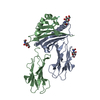
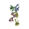
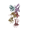
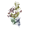
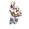
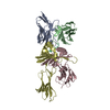
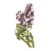
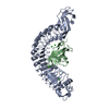
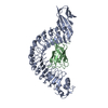
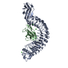
| Title | Crystal structures of immune receptor complexes | ||||||
 Components Components |
| ||||||
 Keywords Keywords | IMMUNE SYSTEM / complex | ||||||
| Function / homology |  Function and homology information Function and homology informationpositive regulation of T cell differentiation / T cell receptor complex / multivesicular body / peptide antigen assembly with MHC class II protein complex / MHC class II protein complex / antigen processing and presentation of exogenous peptide antigen via MHC class II / positive regulation of immune response / peptide antigen binding / positive regulation of T cell activation / MHC class II protein complex binding ...positive regulation of T cell differentiation / T cell receptor complex / multivesicular body / peptide antigen assembly with MHC class II protein complex / MHC class II protein complex / antigen processing and presentation of exogenous peptide antigen via MHC class II / positive regulation of immune response / peptide antigen binding / positive regulation of T cell activation / MHC class II protein complex binding / late endosome membrane / adaptive immune response / early endosome / lysosome / external side of plasma membrane / lysosomal membrane / cell surface / Golgi apparatus Similarity search - Function | ||||||
| Biological species |  | ||||||
| Method |  X-RAY DIFFRACTION / SYNCHROTRON / MOLECULAR REPLACEMENT / Resolution: 2.23 Å X-RAY DIFFRACTION / SYNCHROTRON / MOLECULAR REPLACEMENT / Resolution: 2.23 Å | ||||||
 Authors Authors | Feng, D. / Bond, C.J. / Ely, L.K. / Garcia, K.C. | ||||||
 Citation Citation | Journal: Nat.Immunol. / Year: 2007 Title: Structural evidence for a germline-encoded T cell receptor-major histocompatibility complex interaction 'codon'. Authors: Feng, D. / Bond, C.J. / Ely, L.K. / Maynard, J. / Garcia, K.C. | ||||||
| History |
|
- Structure visualization
| Structure viewer | Molecule: MolmilJmol/JSmol |
|---|
- Downloads & links
-Download
| PDBx/mmCIF format | 2pxy.cif.gz | 140.8 KB | Display | PDBx/mmCIF format |
|---|---|---|---|---|
| PDB format | pdb2pxy.ent.gz | 108.9 KB | Display | PDB format |
| PDBx/mmJSON format | 2pxy.json.gz | Tree view | PDBx/mmJSON format | |
| Others |  Other downloads Other downloads |
-Validation report
| Arichive directory | https://data.pdbj.org/pub/pdb/validation_reports/px/2pxyftp://data.pdbj.org/pub/pdb/validation_reports/px/2pxy | HTTPS FTP |
|---|
-Related structure data


-Links
 PDBj
PDBj


- Assembly
| Deposited unit | 
| ||||||||
|---|---|---|---|---|---|---|---|---|---|
| 1 |
| ||||||||
| Unit cell |
| ||||||||
| Components on special symmetry positions |
|
-Components
-T cell receptor ... , 2 types, 2 molecules AB
| #1: Protein | Mass: 12537.667 Da / Num. of mol.: 1 Source method: isolated from a genetically manipulated source Source: (gene. exp.)  |
|---|---|
| #2: Protein | Mass: 12079.190 Da / Num. of mol.: 1 / Mutation: G17E,H47Y,I75T,L78S Source method: isolated from a genetically manipulated source Source: (gene. exp.) |
-H-2 class II histocompatibility antigen, A-U ... , 2 types, 2 molecules CD
| #3: Protein | Mass: 20771.111 Da / Num. of mol.: 1 / Fragment: extracellular alpha-1, extracellular alpha-2 Source method: isolated from a genetically manipulated source Source: (gene. exp.)  |
|---|---|
| #4: Protein | Mass: 22566.240 Da / Num. of mol.: 1 / Fragment: extracellular beta-1, extracellular beta-2 Source method: isolated from a genetically manipulated source Source: (gene. exp.) |
-Protein/peptide / Non-polymers , 2 types, 330 molecules P
| #5: Protein/peptide | Mass: 1433.511 Da / Num. of mol.: 1 / Mutation: K4Y / Source method: obtained synthetically Details: Synthetic construct. The sequence can be naturally found in Mus musculus (Mouse) |
|---|---|
| #6: Water | ChemComp-HOH / Mass: 18.015 Da / Num. of mol.: 329 / Source method: isolated from a natural source / Formula: H2O |
-Details
| Has protein modification | Y |
|---|
-Experimental details
-Experiment
| Experiment | Method: X-RAY DIFFRACTION / Number of used crystals: 1 |
|---|
- Sample preparation
| Crystal | Density Matthews: 3.01 Å3/Da / Density % sol: 59.12 % |
|---|---|
| Crystal grow | Temperature: 295 K / Method: vapor diffusion, sitting drop / pH: 7 Details: 0.2M Potassium sodium tartrate tetrahydrate, 0.1M succinic acid (pH7.0), 16% polyethylene glycol 3350, VAPOR DIFFUSION, SITTING DROP, temperature 295K |
-Data collection
| Diffraction | Mean temperature: 100 K |
|---|---|
| Diffraction source | Source: SYNCHROTRON / Site: SSRL  / Beamline: BL11-1 / Wavelength: 0.9785 / Beamline: BL11-1 / Wavelength: 0.9785 |
| Detector | Type: ADSC QUANTUM 4 / Detector: CCD / Date: Feb 8, 2006 |
| Radiation | Protocol: SINGLE WAVELENGTH / Monochromatic (M) / Laue (L): M / Scattering type: x-ray |
| Radiation wavelength | Wavelength: 0.9785 Å / Relative weight: 1 |
| Reflection | Resolution: 2.2→47 Å / Num. all: 42263 / Num. obs: 42263 / Redundancy: 4.3 % / Rmerge(I) obs: 0.103 / Rsym value: 0.103 / Net I/σ(I): 23.4 |
| Reflection shell | Resolution: 2.2→2.29 Å / Redundancy: 4.1 % / Rmerge(I) obs: 0.458 / Mean I/σ(I) obs: 3.5 / % possible all: 100 |
- Processing
| Software |
| ||||||||||||||||||||
|---|---|---|---|---|---|---|---|---|---|---|---|---|---|---|---|---|---|---|---|---|---|
| Refinement | Method to determine structure: MOLECULAR REPLACEMENT / Resolution: 2.23→47 Å / σ(F): -1
| ||||||||||||||||||||
| Displacement parameters | Biso mean: 37.2 Å2 | ||||||||||||||||||||
| Refinement step | Cycle: LAST / Resolution: 2.23→47 Å
|
