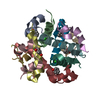
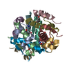
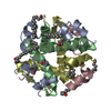
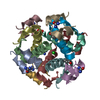
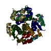
[English] 日本語
Yorodumi
- PDB-2omh: Structure of human insulin cocrystallized with ARG-12 peptide in ... -

+
Open data


ID or keywords:

Loading...

-
Basic information

Entry
Database: PDB / ID: 2omh
TitleStructure of human insulin cocrystallized with ARG-12 peptide in presence of urea
Components
  • Insulin A chain
  • Insulin B chain
KeywordsHORMONE / insulin NPH-like crystal
Function / homology
Function and homology information


negative regulation of NAD(P)H oxidase activity / negative regulation of glycogen catabolic process / regulation of cellular amino acid metabolic process / Signaling by Insulin receptor / IRS activation / nitric oxide-cGMP-mediated signaling / negative regulation of fatty acid metabolic process / Insulin processing / negative regulation of feeding behavior / regulation of protein secretion ...negative regulation of NAD(P)H oxidase activity / negative regulation of glycogen catabolic process / regulation of cellular amino acid metabolic process / Signaling by Insulin receptor / IRS activation / nitric oxide-cGMP-mediated signaling / negative regulation of fatty acid metabolic process / Insulin processing / negative regulation of feeding behavior / regulation of protein secretion / positive regulation of peptide hormone secretion / Regulation of gene expression in beta cells / positive regulation of respiratory burst / positive regulation of dendritic spine maintenance / alpha-beta T cell activation / negative regulation of acute inflammatory response / negative regulation of respiratory burst involved in inflammatory response / negative regulation of protein secretion / fatty acid homeostasis / Synthesis, secretion, and deacylation of Ghrelin / positive regulation of glycogen biosynthetic process / positive regulation of lipid biosynthetic process / Signal attenuation / FOXO-mediated transcription of oxidative stress, metabolic and neuronal genes / negative regulation of gluconeogenesis / positive regulation of nitric oxide mediated signal transduction / regulation of protein localization to plasma membrane / COPI-mediated anterograde transport / negative regulation of lipid catabolic process / negative regulation of oxidative stress-induced intrinsic apoptotic signaling pathway / negative regulation of reactive oxygen species biosynthetic process / positive regulation of insulin receptor signaling pathway / transport vesicle / positive regulation of protein autophosphorylation / Insulin receptor recycling / insulin-like growth factor receptor binding / NPAS4 regulates expression of target genes / positive regulation of protein metabolic process / neuron projection maintenance / endoplasmic reticulum-Golgi intermediate compartment membrane / positive regulation of brown fat cell differentiation / activation of protein kinase B activity / positive regulation of glycolytic process / Insulin receptor signalling cascade / positive regulation of mitotic nuclear division / Regulation of insulin secretion / positive regulation of nitric-oxide synthase activity / positive regulation of long-term synaptic potentiation / endosome lumen / positive regulation of cytokine production / acute-phase response / positive regulation of protein secretion / regulation of transmembrane transporter activity / positive regulation of cell differentiation / positive regulation of glucose import / negative regulation of proteolysis / regulation of synaptic plasticity / wound healing / insulin receptor binding / negative regulation of protein catabolic process / positive regulation of neuron projection development / hormone activity / cognition / Golgi lumen / vasodilation / positive regulation of protein localization to nucleus / glucose metabolic process / regulation of protein localization / glucose homeostasis / cell-cell signaling / insulin receptor signaling pathway / positive regulation of NF-kappaB transcription factor activity / PI5P, PP2A and IER3 Regulate PI3K/AKT Signaling / positive regulation of cell growth / secretory granule lumen / protease binding / positive regulation of MAPK cascade / positive regulation of phosphatidylinositol 3-kinase/protein kinase B signal transduction / positive regulation of cell migration / G protein-coupled receptor signaling pathway / Amyloid fiber formation / endoplasmic reticulum lumen / Golgi membrane / negative regulation of gene expression / positive regulation of cell population proliferation / positive regulation of gene expression / regulation of DNA-templated transcription / extracellular space / extracellular region / identical protein binding
Similarity search - Function
Insulin / Insulin family / Insulin/IGF/Relaxin family / Insulin, conserved site / Insulin family signature. / Insulin-like / Insulin / insulin-like growth factor / relaxin family. / Insulin-like superfamily
Similarity search - Domain/homology
FORMAMIDE / RESORCINOL / UREA / Insulin
Similarity search - Component
Biological speciesHomo sapiens (human)
MethodX-RAY DIFFRACTION / SYNCHROTRON / MOLECULAR REPLACEMENT / Resolution: 1.36 Å
AuthorsNorrman, M. / Schluckebier, G.
CitationJournal: EUR.J.PHARM.SCI. / Year: 2007
Title: Structural characterization of insulin NPH formulations.
Authors: Norrman, M. / Hubalek, F. / Schluckebier, G.
History
DepositionJan 22, 2007Deposition site: RCSB / Processing site: RCSB
Revision 1.0Mar 27, 2007Provider: repository / Type: Initial release
Revision 1.1May 1, 2008Group: Version format compliance
Revision 1.2Jul 13, 2011Group: Advisory / Derived calculations / Version format compliance
Revision 1.3Mar 7, 2018Group: Data collection / Category: diffrn_source / Item: _diffrn_source.pdbx_synchrotron_site
Revision 1.4Dec 27, 2023Group: Data collection / Database references / Derived calculations
Category: chem_comp_atom / chem_comp_bond ...chem_comp_atom / chem_comp_bond / database_2 / pdbx_struct_conn_angle / struct_conn / struct_site
Item: _database_2.pdbx_DOI / _database_2.pdbx_database_accession ..._database_2.pdbx_DOI / _database_2.pdbx_database_accession / _pdbx_struct_conn_angle.ptnr1_auth_asym_id / _pdbx_struct_conn_angle.ptnr1_auth_comp_id / _pdbx_struct_conn_angle.ptnr1_auth_seq_id / _pdbx_struct_conn_angle.ptnr1_label_asym_id / _pdbx_struct_conn_angle.ptnr1_label_atom_id / _pdbx_struct_conn_angle.ptnr1_label_comp_id / _pdbx_struct_conn_angle.ptnr1_label_seq_id / _pdbx_struct_conn_angle.ptnr3_auth_asym_id / _pdbx_struct_conn_angle.ptnr3_auth_comp_id / _pdbx_struct_conn_angle.ptnr3_auth_seq_id / _pdbx_struct_conn_angle.ptnr3_label_asym_id / _pdbx_struct_conn_angle.ptnr3_label_atom_id / _pdbx_struct_conn_angle.ptnr3_label_comp_id / _pdbx_struct_conn_angle.ptnr3_label_seq_id / _pdbx_struct_conn_angle.value / _struct_conn.pdbx_dist_value / _struct_conn.pdbx_leaving_atom_flag / _struct_conn.ptnr1_auth_asym_id / _struct_conn.ptnr1_auth_comp_id / _struct_conn.ptnr1_auth_seq_id / _struct_conn.ptnr1_label_asym_id / _struct_conn.ptnr1_label_atom_id / _struct_conn.ptnr1_label_comp_id / _struct_conn.ptnr1_label_seq_id / _struct_conn.ptnr2_auth_asym_id / _struct_conn.ptnr2_auth_comp_id / _struct_conn.ptnr2_auth_seq_id / _struct_conn.ptnr2_label_asym_id / _struct_conn.ptnr2_label_atom_id / _struct_conn.ptnr2_label_comp_id / _struct_conn.ptnr2_label_seq_id / _struct_site.pdbx_auth_asym_id / _struct_site.pdbx_auth_comp_id / _struct_site.pdbx_auth_seq_id
Revision 1.5Apr 3, 2024Group: Refinement description / Category: pdbx_initial_refinement_model

-
Structure visualization

Structure viewerMolecule:
MolmilJmol/JSmol

Downloads & links

-
Assembly

Deposited unit
A: Insulin A chain
B: Insulin B chain
C: Insulin A chain
D: Insulin B chain
E: Insulin A chain
F: Insulin B chain
hetero molecules


Theoretical massNumber of molelcules
Total (without water)18,23718
Polymers17,4536
Non-polymers78412
Water2,972165
1
A: Insulin A chain
B: Insulin B chain
C: Insulin A chain
D: Insulin B chain
E: Insulin A chain
F: Insulin B chain
hetero molecules

A: Insulin A chain
B: Insulin B chain
C: Insulin A chain
D: Insulin B chain
E: Insulin A chain
F: Insulin B chain
hetero molecules


Theoretical massNumber of molelcules
Total (without water)36,47536
Polymers34,90612
Non-polymers1,56924
Water21612
TypeNameSymmetry operationNumber
identity operation1_555x,y,z1
crystal symmetry operation8_555-y,-x,-z+1/21
Buried area21000 Å2
ΔGint-245 kcal/mol
Surface area13230 Å2
MethodPISA
2
A: Insulin A chain
B: Insulin B chain
C: Insulin A chain
D: Insulin B chain
E: Insulin A chain
F: Insulin B chain
hetero molecules

A: Insulin A chain
B: Insulin B chain
C: Insulin A chain
D: Insulin B chain
E: Insulin A chain
F: Insulin B chain
hetero molecules


Theoretical massNumber of molelcules
Total (without water)36,47536
Polymers34,90612
Non-polymers1,56924
Water21612
TypeNameSymmetry operationNumber
identity operation1_555x,y,z1
crystal symmetry operation7_555y,x,-z1
MethodPQS
Unit cell
Length a, b, c (Å)61.360, 61.360, 85.600
Angle α, β, γ (deg.)90.00, 90.00, 90.00
Int Tables number96
Space group name H-MP43212

-
Components

-
Protein/peptide , 2 types, 6 molecules ACEBDF

#1: Protein/peptide Insulin A chain


Mass: 2383.698 Da / Num. of mol.: 3 / Source method: isolated from a natural source / Source: (natural) Homo sapiens (human) / References: UniProt: P01308
#2: Protein/peptide Insulin B chain


Mass: 3433.953 Da / Num. of mol.: 3 / Source method: isolated from a natural source / Source: (natural) Homo sapiens (human) / References: UniProt: P01308

-
Non-polymers , 7 types, 177 molecules

#3: Chemical ChemComp-NA / SODIUM ION


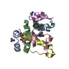
Mass: 22.990 Da / Num. of mol.: 1 / Source method: obtained synthetically / Formula: Na
#4: Chemical ChemComp-RCO / RESORCINOL / 1,3-BENZENEDIOL / 1,3-DIHYDROXYBENZENE / Resorcinol


Mass: 110.111 Da / Num. of mol.: 3 / Source method: obtained synthetically / Formula: C6H6O2
#5: Chemical
ChemComp-URE / UREA / Urea


Mass: 60.055 Da / Num. of mol.: 4 / Source method: obtained synthetically / Formula: CH4N2O
#6: Chemical ChemComp-ZN / ZINC ION


Mass: 65.409 Da / Num. of mol.: 1 / Source method: obtained synthetically / Formula: Zn
#7: Chemical ChemComp-CL / CHLORIDE ION / Chloride


Mass: 35.453 Da / Num. of mol.: 1 / Source method: obtained synthetically / Formula: Cl
#8: Chemical ChemComp-ARF / FORMAMIDE / Formamide


Type: L-peptide NH3 amino terminus / Mass: 45.041 Da / Num. of mol.: 2 / Source method: obtained synthetically / Formula: CH3NO
#9: Water ChemComp-HOH / water / Water


Mass: 18.015 Da / Num. of mol.: 165 / Source method: isolated from a natural source / Formula: H2O

-
Experimental details

-
Experiment

ExperimentMethod: X-RAY DIFFRACTION / Number of used crystals: 1

-
Sample preparation

CrystalDensity Matthews: 2.31 Å3/Da / Density % sol: 46.7 %
Crystal growTemperature: 291 K / Method: vapor diffusion, hanging drop / pH: 7.3
Details: 500mM NaCl, 2.5M urea, 1.2mg/ml ARG-12 peptide, 50mM resorcinol, 50mM phosphate buffer, pH 7.3, VAPOR DIFFUSION, HANGING DROP, temperature 291K

-
Data collection

DiffractionMean temperature: 100 K
Diffraction sourceSource: SYNCHROTRON / Site: MAX II / Beamline: I911-2 / Wavelength: 1 Å
DetectorType: MARMOSAIC 225 mm CCD / Detector: CCD / Date: Oct 7, 2004
RadiationMonochromator: Double crystal monochromator Si(111) / Protocol: SINGLE WAVELENGTH / Monochromatic (M) / Laue (L): M / Scattering type: x-ray
Radiation wavelengthWavelength: 1 Å / Relative weight: 1
ReflectionResolution: 1.36→50 Å / Num. all: 35834 / Num. obs: 34738 / % possible obs: 96.9 % / Redundancy: 5.3 % / Biso Wilson estimate: 24.3 Å2 / Rmerge(I) obs: 0.058 / Net I/σ(I): 12.3
Reflection shellResolution: 1.36→1.4 Å / Redundancy: 4.2 % / Rmerge(I) obs: 0.61 / Mean I/σ(I) obs: 2.2 / Num. unique all: 2933 / % possible all: 98.8

-
Processing

Software
NameVersionClassification
REFMAC5.2.0019refinement
XDSdata reduction
XSCALEdata scaling
MOLREPphasing
RefinementMethod to determine structure: MOLECULAR REPLACEMENT
Starting model: insulin trimer R conformation

Resolution: 1.36→14.95 Å / Cor.coef. Fo:Fc: 0.96 / Cor.coef. Fo:Fc free: 0.952 / SU B: 1.6 / SU ML: 0.038 / TLS residual ADP flag: LIKELY RESIDUAL / Isotropic thermal model: isotropic / Cross valid method: THROUGHOUT / ESU R: 0.063 / ESU R Free: 0.064 / Stereochemistry target values: MAXIMUM LIKELIHOOD / Details: HYDROGENS HAVE BEEN ADDED IN THE RIDING POSITIONS
RfactorNum. reflection% reflectionSelection details
Rfree0.22274 1739 5 %RANDOM
Rwork0.20022 ---
obs0.20136 32948 96.97 %-
Solvent computationIon probe radii: 0.8 Å / Shrinkage radii: 0.8 Å / VDW probe radii: 1.4 Å / Solvent model: MASK
Displacement parametersBiso mean: 22.684 Å2
Baniso -1Baniso -2Baniso -3
1-0.01 Å20 Å20 Å2
2--0.01 Å20 Å2
3----0.03 Å2
Refinement stepCycle: LAST / Resolution: 1.36→14.95 Å
ProteinNucleic acidLigandSolventTotal
Num. atoms1163 0 49 165 1377
Refine LS restraints
Refine-IDTypeDev idealDev ideal targetNumber
X-RAY DIFFRACTIONr_bond_refined_d0.0080.0221253
X-RAY DIFFRACTIONr_angle_refined_deg1.1121.9721697
X-RAY DIFFRACTIONr_dihedral_angle_1_deg5.375147
X-RAY DIFFRACTIONr_dihedral_angle_2_deg32.38924.5961
X-RAY DIFFRACTIONr_dihedral_angle_3_deg11.40215184
X-RAY DIFFRACTIONr_dihedral_angle_4_deg6.699153
X-RAY DIFFRACTIONr_chiral_restr0.0910.2182
X-RAY DIFFRACTIONr_gen_planes_refined0.0040.02970
X-RAY DIFFRACTIONr_nbd_refined0.2330.2603
X-RAY DIFFRACTIONr_nbtor_refined0.3110.2885
X-RAY DIFFRACTIONr_xyhbond_nbd_refined0.1680.290
X-RAY DIFFRACTIONr_metal_ion_refined0.1830.26
X-RAY DIFFRACTIONr_symmetry_vdw_refined0.220.293
X-RAY DIFFRACTIONr_symmetry_hbond_refined0.2090.226
X-RAY DIFFRACTIONr_mcbond_it0.6561.5747
X-RAY DIFFRACTIONr_mcangle_it1.29121186
X-RAY DIFFRACTIONr_scbond_it1.8273521
X-RAY DIFFRACTIONr_scangle_it2.9114.5508
LS refinement shellResolution: 1.36→1.395 Å / Total num. of bins used: 20
RfactorNum. reflection% reflection
Rfree0.35 124 -
Rwork0.308 2414 -
obs--98.79 %
Refinement TLS params.

Method: refined / Refine-ID: X-RAY DIFFRACTION

IDL112)L122)L132)L222)L232)L332)S11 (Å °)S12 (Å °)S13 (Å °)S21 (Å °)S22 (Å °)S23 (Å °)S31 (Å °)S32 (Å °)S33 (Å °)T112)T122)T132)T222)T232)T332)Origin x (Å)Origin y (Å)Origin z (Å)
11.6737-0.34520.6651.2117-0.1261.2633-0.06140.08330.0944-0.0680.05690.0091-0.0561-0.12980.0045-0.0330.02360.00440.00270.0317-0.0215-11.819611.716311.1571
21.181-0.2569-0.4151.3241-0.4272.46320.0656-0.05850.1240.15480.0198-0.0982-0.32230.2368-0.08540.0038-0.05270.0012-0.0047-0.0043-0.01623.675914.845327.772
31.44181.54080.47553.38630.24681.85820.04670.0437-0.2596-0.0796-0.0895-0.39380.22270.24760.0428-0.01640.01740.0411-0.02130.00470.03765.4007-3.18413.2292
Refinement TLS group
IDRefine-IDRefine TLS-IDAuth asym-IDLabel asym-IDAuth seq-IDLabel seq-ID
1X-RAY DIFFRACTION1AA1 - 211 - 21
2X-RAY DIFFRACTION1BB1 - 281 - 28
3X-RAY DIFFRACTION2CC1 - 211 - 21
4X-RAY DIFFRACTION2DD1 - 281 - 28
5X-RAY DIFFRACTION3EE1 - 211 - 21
6X-RAY DIFFRACTION3FF1 - 281 - 28

+
About Yorodumi

-
News

-
Feb 9, 2022. New format data for meta-information of EMDB entries

New format data for meta-information of EMDB entries

  • Version 3 of the EMDB header file is now the official format.
  • The previous official version 1.9 will be removed from the archive.

Related info.:EMDB header

External links:wwPDB to switch to version 3 of the EMDB data model

-
Aug 12, 2020. Covid-19 info

Covid-19 info

URL: https://pdbj.org/emnavi/covid19.php

New page: Covid-19 featured information page in EM Navigator.

Related info.:Covid-19 info / Mar 5, 2020. Novel coronavirus structure data

+
Mar 5, 2020. Novel coronavirus structure data

Novel coronavirus structure data

Related info.:Yorodumi Speices / Aug 12, 2020. Covid-19 info

External links:COVID-19 featured content - PDBj / Molecule of the Month (242):Coronavirus Proteases

+
Jan 31, 2019. EMDB accession codes are about to change! (news from PDBe EMDB page)

EMDB accession codes are about to change! (news from PDBe EMDB page)

  • The allocation of 4 digits for EMDB accession codes will soon come to an end. Whilst these codes will remain in use, new EMDB accession codes will include an additional digit and will expand incrementally as the available range of codes is exhausted. The current 4-digit format prefixed with “EMD-” (i.e. EMD-XXXX) will advance to a 5-digit format (i.e. EMD-XXXXX), and so on. It is currently estimated that the 4-digit codes will be depleted around Spring 2019, at which point the 5-digit format will come into force.
  • The EM Navigator/Yorodumi systems omit the EMD- prefix.

Related info.:Q: What is EMD? / ID/Accession-code notation in Yorodumi/EM Navigator

External links:EMDB Accession Codes are Changing Soon! / Contact to PDBj

+
Jul 12, 2017. Major update of PDB

Major update of PDB

  • wwPDB released updated PDB data conforming to the new PDBx/mmCIF dictionary.
  • This is a major update changing the version number from 4 to 5, and with Remediation, in which all the entries are updated.
  • In this update, many items about electron microscopy experimental information are reorganized (e.g. em_software).
  • Now, EM Navigator and Yorodumi are based on the updated data.

External links:wwPDB Remediation / Enriched Model Files Conforming to OneDep Data Standards Now Available in the PDB FTP Archive

-
Yorodumi

Thousand views of thousand structures

  • Yorodumi is a browser for structure data from EMDB, PDB, SASBDB, etc.
  • This page is also the successor to EM Navigator detail page, and also detail information page/front-end page for Omokage search.
  • The word "yorodu" (or yorozu) is an old Japanese word meaning "ten thousand". "mi" (miru) is to see.

Related info.:EMDB / PDB / SASBDB / Comparison of 3 databanks / Yorodumi Search / Aug 31, 2016. New EM Navigator & Yorodumi / Yorodumi Papers / Jmol/JSmol / Function and homology information / Changes in new EM Navigator and Yorodumi

Read more