 Movie
Movie Controller

[English] 日本語
 Yorodumi
- PDB-2oif: The crystal structure of ferric cyanide bound barley hexacoordina... -
+ Open data

- Basic information
| Entry | Database: PDB / ID: 2oif | ||||||
|---|---|---|---|---|---|---|---|
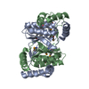
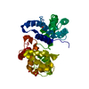
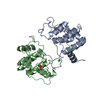
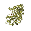
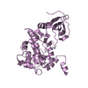
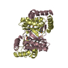
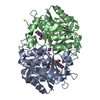
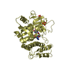
| Title | The crystal structure of ferric cyanide bound barley hexacoordinate hemoglobin. | ||||||
 Components Components | Non-legume hemoglobin | ||||||
 Keywords Keywords | METAL BINDING PROTEIN / hexacoordinate hemoglobin / barley / ligand binding / non-symbiotic / symbiotic / evolution / conformational changes / oxygen transport | ||||||
| Function / homology |  Function and homology information Function and homology informationOxidoreductases; Acting on other nitrogenous compounds as donors; With a cytochrome as acceptor / oxygen binding / oxidoreductase activity / heme binding / metal ion binding / nucleus / cytoplasm Similarity search - Function | ||||||
| Biological species |  | ||||||
| Method |  X-RAY DIFFRACTION / SAD / Resolution: 1.8 Å X-RAY DIFFRACTION / SAD / Resolution: 1.8 Å | ||||||
 Authors Authors | Hoy, J.A. | ||||||
 Citation Citation | Journal: J.Mol.Biol. / Year: 2007 Title: Plant hemoglobins: a molecular fossil record for the evolution of oxygen transport Authors: Hoy, J.A. / Robinson, H. / Trent III, J.T. / Kakar, S. / Smagghe, B.J. / Hargrove, M.S. | ||||||
| History |
|
- Structure visualization
| Structure viewer | Molecule: MolmilJmol/JSmol |
|---|
- Downloads & links
-Download
| PDBx/mmCIF format | 2oif.cif.gz | 276.3 KB | Display | PDBx/mmCIF format |
|---|---|---|---|---|
| PDB format | pdb2oif.ent.gz | 225.7 KB | Display | PDB format |
| PDBx/mmJSON format | 2oif.json.gz | Tree view | PDBx/mmJSON format | |
| Others |  Other downloads Other downloads |
-Validation report
| Arichive directory | https://data.pdbj.org/pub/pdb/validation_reports/oi/2oifftp://data.pdbj.org/pub/pdb/validation_reports/oi/2oif | HTTPS FTP |
|---|
-Related structure data
| Similar structure data |
|---|
-Links
 PDBj
PDBj


- Assembly
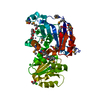
| Deposited unit | 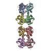
| ||||||||
|---|---|---|---|---|---|---|---|---|---|
| 1 | 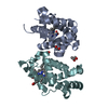
| ||||||||
| 2 | 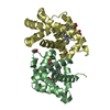
| ||||||||
| 3 | 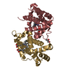
| ||||||||
| 4 | 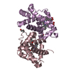
| ||||||||
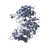
| Unit cell |
|
-Components
| #1: Protein | Mass: 18068.020 Da / Num. of mol.: 8 Source method: isolated from a genetically manipulated source Source: (gene. exp.)  #2: Chemical | ChemComp-CYN /   Mass: 26.017 Da / Num. of mol.: 8 / Source method: obtained synthetically / Formula: CN Mass: 26.017 Da / Num. of mol.: 8 / Source method: obtained synthetically / Formula: CN#3: Chemical | ChemComp-HEM /   Mass: 616.487 Da / Num. of mol.: 8 / Source method: obtained synthetically / Formula: C34H32FeN4O4 Mass: 616.487 Da / Num. of mol.: 8 / Source method: obtained synthetically / Formula: C34H32FeN4O4#4: Chemical | ChemComp-PGO /   Mass: 76.094 Da / Num. of mol.: 13 / Source method: obtained synthetically / Formula: C3H8O2 Mass: 76.094 Da / Num. of mol.: 13 / Source method: obtained synthetically / Formula: C3H8O2#5: Water | ChemComp-HOH / |  Mass: 18.015 Da / Num. of mol.: 876 / Source method: isolated from a natural source / Formula: H2O Mass: 18.015 Da / Num. of mol.: 876 / Source method: isolated from a natural source / Formula: H2O |
|---|
-Experimental details
-Experiment
| Experiment | Method: X-RAY DIFFRACTION / Number of used crystals: 1 |
|---|
- Sample preparation
| Crystal | Density Matthews: 2.73 Å3/Da / Density % sol: 54.97 % |
|---|---|
| Crystal grow | Temperature: 277 K / Method: vapor diffusion, hanging drop / pH: 5.3 Details: 20% PEG 3350, 0.2 M ammonium citrate, 4% polypropylene glycol, 0.1 M sodium cyanide, 0.01 M hepes, pH 5.3, VAPOR DIFFUSION, HANGING DROP, temperature 277K |
-Data collection
| Diffraction | Mean temperature: 100 K |
|---|---|
| Diffraction source | Source: ROTATING ANODE / Type: RIGAKU RU200 / Wavelength: 1.5418 Å |
| Detector | Type: RIGAKU RAXIS IV / Detector: IMAGE PLATE / Date: Apr 1, 2006 |
| Radiation | Protocol: SINGLE WAVELENGTH / Monochromatic (M) / Laue (L): M / Scattering type: x-ray |
| Radiation wavelength | Wavelength: 1.5418 Å / Relative weight: 1 |
| Reflection | Resolution: 1.8→30 Å / Num. all: 141630 / Num. obs: 123785 / % possible obs: 87.4 % / Redundancy: 3.7 % / Rmerge(I) obs: 0.045 |
| Reflection shell | Resolution: 1.8→1.86 Å / Redundancy: 2.6 % / Rmerge(I) obs: 0.378 / % possible all: 47.8 |
- Processing
| Software |
| ||||||||||||||||||||||||||||||||||||||||||||||||||||||||||||||||||||||||||||||||||||||||||||||||||||||||||||||||||||||||||||||||||||||||
|---|---|---|---|---|---|---|---|---|---|---|---|---|---|---|---|---|---|---|---|---|---|---|---|---|---|---|---|---|---|---|---|---|---|---|---|---|---|---|---|---|---|---|---|---|---|---|---|---|---|---|---|---|---|---|---|---|---|---|---|---|---|---|---|---|---|---|---|---|---|---|---|---|---|---|---|---|---|---|---|---|---|---|---|---|---|---|---|---|---|---|---|---|---|---|---|---|---|---|---|---|---|---|---|---|---|---|---|---|---|---|---|---|---|---|---|---|---|---|---|---|---|---|---|---|---|---|---|---|---|---|---|---|---|---|---|---|---|
| Refinement | Method to determine structure: SAD / Resolution: 1.8→30 Å / Cor.coef. Fo:Fc: 0.958 / Cor.coef. Fo:Fc free: 0.936 / Cross valid method: THROUGHOUT / σ(F): 0 / Stereochemistry target values: MAXIMUM LIKELIHOOD
| ||||||||||||||||||||||||||||||||||||||||||||||||||||||||||||||||||||||||||||||||||||||||||||||||||||||||||||||||||||||||||||||||||||||||
| Solvent computation | Ion probe radii: 0.8 Å / Shrinkage radii: 0.8 Å / VDW probe radii: 1.4 Å / Solvent model: BABINET MODEL WITH MASK | ||||||||||||||||||||||||||||||||||||||||||||||||||||||||||||||||||||||||||||||||||||||||||||||||||||||||||||||||||||||||||||||||||||||||
| Displacement parameters | Biso mean: 35.119 Å2
| ||||||||||||||||||||||||||||||||||||||||||||||||||||||||||||||||||||||||||||||||||||||||||||||||||||||||||||||||||||||||||||||||||||||||
| Refinement step | Cycle: LAST / Resolution: 1.8→30 Å
| ||||||||||||||||||||||||||||||||||||||||||||||||||||||||||||||||||||||||||||||||||||||||||||||||||||||||||||||||||||||||||||||||||||||||
| Refine LS restraints |
| ||||||||||||||||||||||||||||||||||||||||||||||||||||||||||||||||||||||||||||||||||||||||||||||||||||||||||||||||||||||||||||||||||||||||
| LS refinement shell | Resolution: 1.801→1.848 Å / Total num. of bins used: 20 /
|
