- PDB-2odk: Putative prevent-host-death protein from Nitrosomonas europaea -
+
Open data
ID or keywords:
Loading...
-
Basic information
Entry
Database: PDB / ID: 2odk
Title
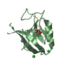
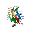
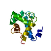
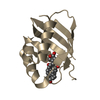
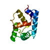
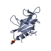
Putative prevent-host-death protein from Nitrosomonas europaea
Components
Hypothetical protein
Keywords
STRUCTURAL GENOMICS / UNKNOWN FUNCTION / prevent-host-death protein / APC7367 / PSI-2 / Protein Structure Initiative / Midwest Center for Structural Genomics / MCSG
Function / homology
YefM-like domain / Type II toxin-antitoxin system, antitoxin Phd/YefM / Antitoxin Phd_YefM, type II toxin-antitoxin system / YefM-like superfamily / YefM-like fold / 3-Layer(aba) Sandwich / Alpha Beta / Antitoxin
Function and homology information
Biological species
Nitrosomonas europaea (bacteria)
Method
X-RAY DIFFRACTION / SYNCHROTRON / SAD / Resolution: 1.4 Å
BIOMOLECULE: 1, 2 THIS ENTRY CONTAINS THE CRYSTALLOGRAPHIC ASYMMETRIC UNIT WHICH CONSISTS OF 4 ... BIOMOLECULE: 1, 2 THIS ENTRY CONTAINS THE CRYSTALLOGRAPHIC ASYMMETRIC UNIT WHICH CONSISTS OF 4 CHAIN(S). SEE REMARK 350 FOR INFORMATION ON GENERATING THE BIOLOGICAL MOLECULE(S). AUTHORS STATE THAT THE DIMER IS A PUTATIVE BIOLOGICAL UNIT BASED ON ANALYSIS OF THE PROTEIN INTERFACES.
Remark 999
SEQUENCE AUTHORS STATE THAT THE C-TERMINUS OF THIS PROTEIN IS FLEXIBLE AND THEREFORE THE RESIDUES ... SEQUENCE AUTHORS STATE THAT THE C-TERMINUS OF THIS PROTEIN IS FLEXIBLE AND THEREFORE THE RESIDUES 52-87 ARE MISSING IN COORDINATES DUE TO LACK OF ELECTRON DENSITY.
Resolution: 1.4→1.43 Å / Redundancy: 3.1 % / Rmerge(I) obs: 0.377 / Mean I/σ(I) obs: 2.03 / % possible all: 62.7
-
Processing
Software
Name
Version
Classification
REFMAC
5.2.0019
refinement
SBC-Collect
datacollection
HKL-3000
datareduction
HKL-3000
datascaling
SHELXD
phasing
MLPHARE
phasing
DM
phasing
SOLVE
phasing
RESOLVE
phasing
HKL-3000
phasing
Refinement
Method to determine structure: SAD / Resolution: 1.4→37.1 Å / Cor.coef. Fo:Fc: 0.976 / Cor.coef. Fo:Fc free: 0.971 / SU B: 1.341 / SU ML: 0.024 / Cross valid method: THROUGHOUT / σ(F): 0 / ESU R: 0.048 / ESU R Free: 0.044 / Stereochemistry target values: MAXIMUM LIKELIHOOD / Details: HYDROGENS HAVE BEEN ADDED IN THE RIDING POSITIONS
Rfactor
Num. reflection
% reflection
Selection details
Rfree
0.15379
2838
5.1 %
RANDOM
Rwork
0.13386
-
-
-
obs
0.13487
53222
96.34 %
-
all
-
56060
-
-
Solvent computation
Ion probe radii: 0.8 Å / Shrinkage radii: 0.8 Å / VDW probe radii: 1.2 Å / Solvent model: MASK
Displacement parameters
Biso mean: 18.243 Å2
Baniso -1
Baniso -2
Baniso -3
1-
0.4 Å2
0 Å2
0.02 Å2
2-
-
0.03 Å2
0 Å2
3-
-
-
-0.41 Å2
Refinement step
Cycle: LAST / Resolution: 1.4→37.1 Å
Protein
Nucleic acid
Ligand
Solvent
Total
Num. atoms
1606
0
16
312
1934
Refine LS restraints
Refine-ID
Type
Dev ideal
Dev ideal target
Number
X-RAY DIFFRACTION
r_bond_refined_d
0.015
0.022
1885
X-RAY DIFFRACTION
r_bond_other_d
X-RAY DIFFRACTION
r_angle_refined_deg
1.616
1.949
2607
X-RAY DIFFRACTION
r_angle_other_deg
X-RAY DIFFRACTION
r_dihedral_angle_1_deg
5.322
5
263
X-RAY DIFFRACTION
r_dihedral_angle_2_deg
29.389
23.107
103
X-RAY DIFFRACTION
r_dihedral_angle_3_deg
11.91
15
329
X-RAY DIFFRACTION
r_dihedral_angle_4_deg
16.121
15
25
X-RAY DIFFRACTION
r_chiral_restr
0.121
0.2
283
X-RAY DIFFRACTION
r_gen_planes_refined
0.008
0.02
1506
X-RAY DIFFRACTION
r_gen_planes_other
X-RAY DIFFRACTION
r_nbd_refined
0.227
0.2
966
X-RAY DIFFRACTION
r_nbd_other
X-RAY DIFFRACTION
r_nbtor_refined
0.315
0.2
1289
X-RAY DIFFRACTION
r_nbtor_other
X-RAY DIFFRACTION
r_xyhbond_nbd_refined
0.186
0.2
250
X-RAY DIFFRACTION
r_xyhbond_nbd_other
X-RAY DIFFRACTION
r_metal_ion_refined
X-RAY DIFFRACTION
r_metal_ion_other
X-RAY DIFFRACTION
r_symmetry_vdw_refined
0.455
0.2
59
X-RAY DIFFRACTION
r_symmetry_vdw_other
X-RAY DIFFRACTION
r_symmetry_hbond_refined
0.256
0.2
41
X-RAY DIFFRACTION
r_symmetry_hbond_other
X-RAY DIFFRACTION
r_symmetry_metal_ion_refined
X-RAY DIFFRACTION
r_symmetry_metal_ion_other
X-RAY DIFFRACTION
r_mcbond_it
1.792
1.5
1162
X-RAY DIFFRACTION
r_mcbond_other
X-RAY DIFFRACTION
r_mcangle_it
2.535
2
1837
X-RAY DIFFRACTION
r_scbond_it
3.452
3
830
X-RAY DIFFRACTION
r_scangle_it
4.845
4.5
737
X-RAY DIFFRACTION
r_rigid_bond_restr
2.168
3
1992
X-RAY DIFFRACTION
r_sphericity_free
7.048
3
313
X-RAY DIFFRACTION
r_sphericity_bonded
4.829
3
1817
LS refinement shell
Resolution: 1.4→1.436 Å / Total num. of bins used: 20
Rfactor
Num. reflection
% reflection
Rfree
0.304
141
-
Rwork
0.24
2692
-
obs
-
-
65.7 %
+
About Yorodumi
-
News
-
Feb 9, 2022. New format data for meta-information of EMDB entries
New format data for meta-information of EMDB entries
Version 3 of the EMDB header file is now the official format.
The previous official version 1.9 will be removed from the archive.
In the structure databanks used in Yorodumi, some data are registered as the other names, "COVID-19 virus" and "2019-nCoV". Here are the details of the virus and the list of structure data.
Jan 31, 2019. EMDB accession codes are about to change! (news from PDBe EMDB page)
EMDB accession codes are about to change! (news from PDBe EMDB page)
The allocation of 4 digits for EMDB accession codes will soon come to an end. Whilst these codes will remain in use, new EMDB accession codes will include an additional digit and will expand incrementally as the available range of codes is exhausted. The current 4-digit format prefixed with “EMD-” (i.e. EMD-XXXX) will advance to a 5-digit format (i.e. EMD-XXXXX), and so on. It is currently estimated that the 4-digit codes will be depleted around Spring 2019, at which point the 5-digit format will come into force.
The EM Navigator/Yorodumi systems omit the EMD- prefix.
Related info.:Q: What is EMD? / ID/Accession-code notation in Yorodumi/EM Navigator
Yorodumi is a browser for structure data from EMDB, PDB, SASBDB, etc.
This page is also the successor to EM Navigator detail page, and also detail information page/front-end page for Omokage search.
The word "yorodu" (or yorozu) is an old Japanese word meaning "ten thousand". "mi" (miru) is to see.
Related info.:EMDB / PDB / SASBDB / Comparison of 3 databanks / Yorodumi Search / Aug 31, 2016. New EM Navigator & Yorodumi / Yorodumi Papers / Jmol/JSmol / Function and homology information / Changes in new EM Navigator and Yorodumi
 Movie
Movie Controller


 Open data
 Basic information
 Components
 Keywords
 Function and homology information
 Nitrosomonas europaea (bacteria)
 X-RAY DIFFRACTION /
  Authors
 Citation
 Structure visualization
 Downloads & links
 Other downloads


 PDBj
PDBj Assembly


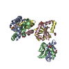

 Mass: 96.063 Da / Num. of mol.: 2 / Source method: obtained synthetically / Formula: SO4
 Mass: 92.094 Da / Num. of mol.: 1 / Source method: obtained synthetically / Formula: C3H8O3
 Mass: 18.015 Da / Num. of mol.: 312 / Source method: isolated from a natural source / Formula: H2O
 Sample preparation
 / Beamline: 19-ID / Wavelength: 0.979 Å
 Processing