 Movie
Movie Controller


+ Open data

- Basic information
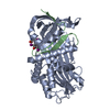
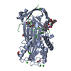
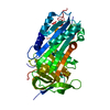
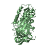
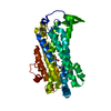
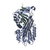
| Entry | Database: PDB / ID: 2oay | ||||||
|---|---|---|---|---|---|---|---|
| Title | Crystal structure of latent human C1-inhibitor | ||||||
 Components Components | Plasma protease C1 inhibitor | ||||||
 Keywords Keywords | IMMUNE SYSTEM / HYDROLASE INHIBITOR / LATENT SERPIN / RCL INSERTION | ||||||
| Function / homology |  Function and homology information Function and homology informationnegative regulation of complement activation, lectin pathway / Defective SERPING1 causes hereditary angioedema / blood circulation / fibrinolysis / : / platelet alpha granule lumen / Regulation of Complement cascade / serine-type endopeptidase inhibitor activity / blood coagulation / Platelet degranulation ...negative regulation of complement activation, lectin pathway / Defective SERPING1 causes hereditary angioedema / blood circulation / fibrinolysis / : / platelet alpha granule lumen / Regulation of Complement cascade / serine-type endopeptidase inhibitor activity / blood coagulation / Platelet degranulation / extracellular matrix / blood microparticle / endoplasmic reticulum lumen / : / extracellular exosome / extracellular region Similarity search - Function | ||||||
| Biological species |  Homo sapiens (human) Homo sapiens (human) | ||||||
| Method |  X-RAY DIFFRACTION / SYNCHROTRON / MOLECULAR REPLACEMENT / Resolution: 2.35 Å X-RAY DIFFRACTION / SYNCHROTRON / MOLECULAR REPLACEMENT / Resolution: 2.35 Å | ||||||
 Authors Authors | Harmat, V. / Beinrohr, L. / Gal, P. / Dobo, J. | ||||||
 Citation Citation | Journal: J.Biol.Chem. / Year: 2007 Title: C1 inhibitor serpin domain structure reveals the likely mechanism of heparin potentiation and conformational disease Authors: Beinrohr, L. / Harmat, V. / Dobo, J. / Lorincz, Z. / Gal, P. / Zavodszky, P. | ||||||
| History |
|
- Structure visualization
| Structure viewer | Molecule: MolmilJmol/JSmol |
|---|
- Downloads & links
-Download
| PDBx/mmCIF format | 2oay.cif.gz | 89.2 KB | Display | PDBx/mmCIF format |
|---|---|---|---|---|
| PDB format | pdb2oay.ent.gz | 65.8 KB | Display | PDB format |
| PDBx/mmJSON format | 2oay.json.gz | Tree view | PDBx/mmJSON format | |
| Others |  Other downloads Other downloads |
-Validation report
| Arichive directory | https://data.pdbj.org/pub/pdb/validation_reports/oa/2oayftp://data.pdbj.org/pub/pdb/validation_reports/oa/2oay | HTTPS FTP |
|---|
-Related structure data
| Related structure data |  1c8oS  1dvmS  1e05S 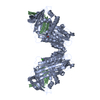 1hleS 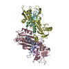 1jjoS  1jrrS 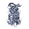 1jtiS 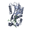 1mtpS  1qmbS  4caaS S: Starting model for refinement |
|---|---|
| Similar structure data |


-Links
 PDBj
PDBj


- Assembly
| Deposited unit | 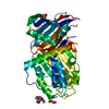
| ||||||||
|---|---|---|---|---|---|---|---|---|---|
| 1 |
| ||||||||
| Unit cell |
|
-Components
| #1: Protein | Mass: 43903.484 Da / Num. of mol.: 1 / Fragment: serpin domain / Mutation: allele V458M Source method: isolated from a genetically manipulated source Source: (gene. exp.) Homo sapiens (human) / Gene: SERPING1, C1IN, C1NH / Plasmid: pPic9K / Production host:  Pichia pastoris (fungus) / Strain (production host): GS115 / References: UniProt: P05155 Pichia pastoris (fungus) / Strain (production host): GS115 / References: UniProt: P05155 | ||||
|---|---|---|---|---|---|
| #2: Sugar | ChemComp-NAG /   Type: D-saccharide, beta linking / Mass: 221.208 Da / Num. of mol.: 1 Type: D-saccharide, beta linking / Mass: 221.208 Da / Num. of mol.: 1Source method: isolated from a genetically manipulated source Formula: C8H15NO6 | ||||
| #3: Chemical |   Mass: 92.094 Da / Num. of mol.: 3 / Source method: obtained synthetically / Formula: C3H8O3 Mass: 92.094 Da / Num. of mol.: 3 / Source method: obtained synthetically / Formula: C3H8O3#4: Water | ChemComp-HOH / |  Mass: 18.015 Da / Num. of mol.: 70 / Source method: isolated from a natural source / Formula: H2O Mass: 18.015 Da / Num. of mol.: 70 / Source method: isolated from a natural source / Formula: H2OHas protein modification | Y | |
-Experimental details
-Experiment
| Experiment | Method: X-RAY DIFFRACTION / Number of used crystals: 1 |
|---|
- Sample preparation
| Crystal | Density Matthews: 3.04 Å3/Da / Density % sol: 59.59 % |
|---|---|
| Crystal grow | Temperature: 293 K / Method: vapor diffusion, hanging drop / pH: 7.5 Details: 0.85 M NaH2PO4, 0.85 M KH2PO4, 0.1 M HEPES, pH 7.5, VAPOR DIFFUSION, HANGING DROP, temperature 293K |
-Data collection
| Diffraction | Mean temperature: 100 K |
|---|---|
| Diffraction source | Source: SYNCHROTRON / Site: EMBL/DESY, HAMBURG  / Beamline: X11 / Wavelength: 0.8162 Å / Beamline: X11 / Wavelength: 0.8162 Å |
| Detector | Type: MAR CCD 165 mm / Detector: CCD / Date: May 3, 2006 / Details: mirrors |
| Radiation | Monochromator: Si [111] / Protocol: SINGLE WAVELENGTH / Monochromatic (M) / Laue (L): M / Scattering type: x-ray |
| Radiation wavelength | Wavelength: 0.8162 Å / Relative weight: 1 |
| Reflection | Resolution: 2.35→84.52 Å / Num. obs: 21930 / % possible obs: 99.8 % / Observed criterion σ(I): -3 / Redundancy: 14.6 % / Biso Wilson estimate: 32.4 Å2 / Rmerge(I) obs: 0.097 / Net I/σ(I): 20.29 |
| Reflection shell | Resolution: 2.35→2.4 Å / Redundancy: 9.54 % / Rmerge(I) obs: 0.652 / Mean I/σ(I) obs: 3.89 / Num. unique all: 1341 / % possible all: 100 |
- Processing
| Software |
| ||||||||||||||||||||||||||||||||||||||||||||||||||||||||||||||||||||||||||||||||||||||||||||||||||||||||||||||||||||||||||||||||||||||||||||||||||||||||||||||||||||||||||
|---|---|---|---|---|---|---|---|---|---|---|---|---|---|---|---|---|---|---|---|---|---|---|---|---|---|---|---|---|---|---|---|---|---|---|---|---|---|---|---|---|---|---|---|---|---|---|---|---|---|---|---|---|---|---|---|---|---|---|---|---|---|---|---|---|---|---|---|---|---|---|---|---|---|---|---|---|---|---|---|---|---|---|---|---|---|---|---|---|---|---|---|---|---|---|---|---|---|---|---|---|---|---|---|---|---|---|---|---|---|---|---|---|---|---|---|---|---|---|---|---|---|---|---|---|---|---|---|---|---|---|---|---|---|---|---|---|---|---|---|---|---|---|---|---|---|---|---|---|---|---|---|---|---|---|---|---|---|---|---|---|---|---|---|---|---|---|---|---|---|---|---|
| Refinement | Method to determine structure: MOLECULAR REPLACEMENT Starting model: Ensemble of truncated serpin structures. PDB codes: 4CAA, 1QMB, 1E05, 1DVM, 1C8O, 1JTI, 1JJO, 1MTP, 1HLE, 1JRR Resolution: 2.35→84.52 Å / Cor.coef. Fo:Fc: 0.966 / Cor.coef. Fo:Fc free: 0.948 / SU B: 6.574 / SU ML: 0.154 / TLS residual ADP flag: LIKELY RESIDUAL / Isotropic thermal model: Isotropic / Cross valid method: THROUGHOUT / σ(F): 0 / σ(I): 0 / ESU R: 0.239 / ESU R Free: 0.197 / Stereochemistry target values: MAXIMUM LIKELIHOOD / Details: HYDROGENS HAVE BEEN ADDED IN THE RIDING POSITIONS
| ||||||||||||||||||||||||||||||||||||||||||||||||||||||||||||||||||||||||||||||||||||||||||||||||||||||||||||||||||||||||||||||||||||||||||||||||||||||||||||||||||||||||||
| Solvent computation | Ion probe radii: 0.8 Å / Shrinkage radii: 0.8 Å / VDW probe radii: 1.4 Å / Solvent model: BABINET MODEL WITH MASK | ||||||||||||||||||||||||||||||||||||||||||||||||||||||||||||||||||||||||||||||||||||||||||||||||||||||||||||||||||||||||||||||||||||||||||||||||||||||||||||||||||||||||||
| Displacement parameters | Biso mean: 32.433 Å2
| ||||||||||||||||||||||||||||||||||||||||||||||||||||||||||||||||||||||||||||||||||||||||||||||||||||||||||||||||||||||||||||||||||||||||||||||||||||||||||||||||||||||||||
| Refinement step | Cycle: LAST / Resolution: 2.35→84.52 Å
| ||||||||||||||||||||||||||||||||||||||||||||||||||||||||||||||||||||||||||||||||||||||||||||||||||||||||||||||||||||||||||||||||||||||||||||||||||||||||||||||||||||||||||
| Refine LS restraints |
| ||||||||||||||||||||||||||||||||||||||||||||||||||||||||||||||||||||||||||||||||||||||||||||||||||||||||||||||||||||||||||||||||||||||||||||||||||||||||||||||||||||||||||
| LS refinement shell | Resolution: 2.35→2.411 Å / Total num. of bins used: 20 /
| ||||||||||||||||||||||||||||||||||||||||||||||||||||||||||||||||||||||||||||||||||||||||||||||||||||||||||||||||||||||||||||||||||||||||||||||||||||||||||||||||||||||||||
| Refinement TLS params. | Method: refined / Origin x: -14.7946 Å / Origin y: 71.6738 Å / Origin z: -0.6784 Å
| ||||||||||||||||||||||||||||||||||||||||||||||||||||||||||||||||||||||||||||||||||||||||||||||||||||||||||||||||||||||||||||||||||||||||||||||||||||||||||||||||||||||||||
| Refinement TLS group |
|
