 Movie
Movie Controller

[English] 日本語
 Yorodumi
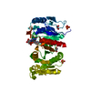
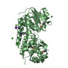
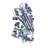
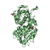
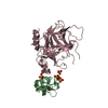
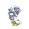
- PDB-1mtp: The X-ray crystal structure of a serpin from a thermophilic prokaryote -
+ Open data

- Basic information
| Entry | Database: PDB / ID: 1mtp | ||||||
|---|---|---|---|---|---|---|---|
| Title | The X-ray crystal structure of a serpin from a thermophilic prokaryote | ||||||
 Components Components |
| ||||||
 Keywords Keywords | STRUCTURAL GENOMICS / protease inhibitor | ||||||
| Function / homology |  Function and homology information Function and homology information | ||||||
| Biological species |   Thermobifida fusca (bacteria) Thermobifida fusca (bacteria) | ||||||
| Method |  X-RAY DIFFRACTION / MOLECULAR REPLACEMENT / Resolution: 1.5 Å X-RAY DIFFRACTION / MOLECULAR REPLACEMENT / Resolution: 1.5 Å | ||||||
 Authors Authors | Irving, J.A. / Cabrita, L.D. / Rossjohn, J. / Pike, R.N. / Bottomley, S.P. / Whisstock, J.C. | ||||||
 Citation Citation | Journal: Structure / Year: 2003 Title: The 1.5 A crystal structure of a prokaryote serpin: controlling conformational change in a heated environment Authors: Irving, J.A. / Cabrita, L.D. / Rossjohn, J. / Pike, R.N. / Bottomley, S.P. / Whisstock, J.C. | ||||||
| History |
|
- Structure visualization
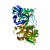
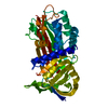
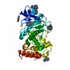
| Structure viewer | Molecule: MolmilJmol/JSmol |
|---|
- Downloads & links
-Download
| PDBx/mmCIF format | 1mtp.cif.gz | 92.5 KB | Display | PDBx/mmCIF format |
|---|---|---|---|---|
| PDB format | pdb1mtp.ent.gz | 69.1 KB | Display | PDB format |
| PDBx/mmJSON format | 1mtp.json.gz | Tree view | PDBx/mmJSON format | |
| Others |  Other downloads Other downloads |
-Validation report
| Arichive directory | https://data.pdbj.org/pub/pdb/validation_reports/mt/1mtpftp://data.pdbj.org/pub/pdb/validation_reports/mt/1mtp | HTTPS FTP |
|---|
-Related structure data
| Related structure data | 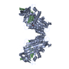 1hleS S: Starting model for refinement |
|---|---|
| Similar structure data |

-Links
 PDBj
PDBj
- Assembly
| Deposited unit | 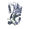
| ||||||||
|---|---|---|---|---|---|---|---|---|---|
| 1 |
| ||||||||
| Unit cell |
|
-Components
| #1: Protein | Mass: 34976.918 Da / Num. of mol.: 1 / Fragment: Chain A, residue 55-377 Source method: isolated from a genetically manipulated source Source: (gene. exp.) Thermobifida fusca (bacteria) / Production host: |
|---|---|
| #2: Protein/peptide | Mass: 4787.580 Da / Num. of mol.: 1 / Fragment: Chain B, residue 378-420 Source method: isolated from a genetically manipulated source Source: (gene. exp.) Thermobifida fusca (bacteria) / Production host: |
| #3: Water | ChemComp-HOH /  Mass: 18.015 Da / Num. of mol.: 604 / Source method: isolated from a natural source / Formula: H2O Mass: 18.015 Da / Num. of mol.: 604 / Source method: isolated from a natural source / Formula: H2O |
-Experimental details
-Experiment
| Experiment | Method: X-RAY DIFFRACTION / Number of used crystals: 1 |
|---|
- Sample preparation
| Crystal | Density Matthews: 2.16 Å3/Da / Density % sol: 42.94 % | ||||||||||||||||||||||||
|---|---|---|---|---|---|---|---|---|---|---|---|---|---|---|---|---|---|---|---|---|---|---|---|---|---|
| Crystal grow | Temperature: 294 K / Method: vapor diffusion, hanging drop / pH: 9 Details: PEG 4000, 0.1 M Tris, pH 9.0, VAPOR DIFFUSION, HANGING DROP, temperature 294K | ||||||||||||||||||||||||
| Crystal grow | *PLUS PH range low: 9 / PH range high: 8 | ||||||||||||||||||||||||
| Components of the solutions | *PLUS
|
-Data collection
| Diffraction | Mean temperature: 100 K |
|---|---|
| Diffraction source | Source: ROTATING ANODE / Type: RIGAKU RUH3R / Wavelength: 1.5418 Å |
| Detector | Type: RIGAKU RAXIS IV / Detector: IMAGE PLATE / Date: Mar 14, 2002 / Details: OSMIC MIRRORS |
| Radiation | Protocol: SINGLE WAVELENGTH / Monochromatic (M) / Laue (L): M / Scattering type: x-ray |
| Radiation wavelength | Wavelength: 1.5418 Å / Relative weight: 1 |
| Reflection | Resolution: 1.5→50 Å / Num. all: 179258 / Num. obs: 179258 / Observed criterion σ(I): 0 |
| Reflection | *PLUS Num. obs: 54979 / % possible obs: 98.1 % / Redundancy: 3.3 % / Num. measured all: 179258 / Rmerge(I) obs: 0.043 |
| Reflection shell | *PLUS % possible obs: 92 % / Rmerge(I) obs: 0.199 / Mean I/σ(I) obs: 2.6 |
- Processing
| Software |
| ||||||||||||||||||||||||
|---|---|---|---|---|---|---|---|---|---|---|---|---|---|---|---|---|---|---|---|---|---|---|---|---|---|
| Refinement | Method to determine structure: MOLECULAR REPLACEMENT Starting model: PDB ENTRY 1HLE Resolution: 1.5→50 Å
| ||||||||||||||||||||||||
| Refinement step | Cycle: LAST / Resolution: 1.5→50 Å
| ||||||||||||||||||||||||
| Refinement | *PLUS Lowest resolution: 50 Å / % reflection Rfree: 3 % | ||||||||||||||||||||||||
| Solvent computation | *PLUS | ||||||||||||||||||||||||
| Displacement parameters | *PLUS | ||||||||||||||||||||||||
| Refine LS restraints | *PLUS
|
