 Movie
Movie Controller

[English] 日本語
 Yorodumi
- PDB-1hle: CRYSTAL STRUCTURE OF CLEAVED EQUINE LEUCOCYTE ELASTASE INHIBITOR ... -
+ Open data

- Basic information
| Entry | Database: PDB / ID: 1hle | ||||||
|---|---|---|---|---|---|---|---|
| Title | CRYSTAL STRUCTURE OF CLEAVED EQUINE LEUCOCYTE ELASTASE INHIBITOR DETERMINED AT 1.95 ANGSTROMS RESOLUTION | ||||||
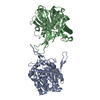
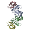
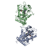
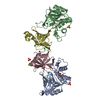
 Components Components | (HORSE LEUKOCYTE ELASTASE INHIBITOR) x 2 | ||||||
 Keywords Keywords | HYDROLASE INHIBITOR(SERINE PROTEINASE) | ||||||
| Function / homology |  Function and homology information Function and homology informationcytolytic granule / negative regulation of interleukin-1 beta production / serine-type endopeptidase inhibitor activity / early endosome / : Similarity search - Function | ||||||
| Biological species |  | ||||||
| Method |  X-RAY DIFFRACTION / Resolution: 1.95 Å X-RAY DIFFRACTION / Resolution: 1.95 Å | ||||||
 Authors Authors | Baumann, U. / Bode, W. / Huber, R. / Travis, J. / Potempa, J. | ||||||
 Citation Citation | Journal: J.Mol.Biol. / Year: 1992 Title: Crystal structure of cleaved equine leucocyte elastase inhibitor determined at 1.95 A resolution. Authors: Baumann, U. / Bode, W. / Huber, R. / Travis, J. / Potempa, J. | ||||||
| History |
|
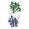
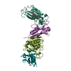
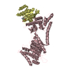
- Structure visualization
| Structure viewer | Molecule: MolmilJmol/JSmol |
|---|
- Downloads & links
-Download
| PDBx/mmCIF format | 1hle.cif.gz | 112.5 KB | Display | PDBx/mmCIF format |
|---|---|---|---|---|
| PDB format | pdb1hle.ent.gz | 86.1 KB | Display | PDB format |
| PDBx/mmJSON format | 1hle.json.gz | Tree view | PDBx/mmJSON format | |
| Others |  Other downloads Other downloads |
-Validation report
| Arichive directory | https://data.pdbj.org/pub/pdb/validation_reports/hl/1hleftp://data.pdbj.org/pub/pdb/validation_reports/hl/1hle | HTTPS FTP |
|---|
-Related structure data
| Similar structure data |
|---|


-Links
 PDBj
PDBj
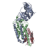
- Assembly
| Deposited unit | 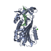
| ||||||||
|---|---|---|---|---|---|---|---|---|---|
| 1 | 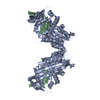
| ||||||||
| Unit cell |
|
-Components
| #1: Protein | Mass: 38957.539 Da / Num. of mol.: 1 Source method: isolated from a genetically manipulated source Source: (gene. exp.) | ||
|---|---|---|---|
| #2: Protein/peptide | Mass: 3639.040 Da / Num. of mol.: 1 Source method: isolated from a genetically manipulated source Source: (gene. exp.) | ||
| #3: Chemical | ChemComp-CA /   Mass: 40.078 Da / Num. of mol.: 1 / Source method: obtained synthetically / Formula: Ca Mass: 40.078 Da / Num. of mol.: 1 / Source method: obtained synthetically / Formula: Ca | ||
| #4: Water | ChemComp-HOH /  Mass: 18.015 Da / Num. of mol.: 212 / Source method: isolated from a natural source / Formula: H2O Mass: 18.015 Da / Num. of mol.: 212 / Source method: isolated from a natural source / Formula: H2O | ||
| Has protein modification | Y | ||
| Nonpolymer details | A CA 2+ ION HAS BEEN IDENTIFIED| Sequence details | THE SEQUENCE USED IS THAT OF DUBIN ET AL., (J. BIOL. CHEM., 1992). BASED ON PEPTIDE SEQUENCING AND ...THE SEQUENCE USED IS THAT OF DUBIN ET AL., (J. BIOL. CHEM., 1992). BASED ON PEPTIDE SEQUENCING | |
-Experimental details
-Experiment
| Experiment | Method: X-RAY DIFFRACTION |
|---|
- Sample preparation
| Crystal | Density Matthews: 2.98 Å3/Da / Density % sol: 58.78 % | ||||||||||||||||||||||||||||||
|---|---|---|---|---|---|---|---|---|---|---|---|---|---|---|---|---|---|---|---|---|---|---|---|---|---|---|---|---|---|---|---|
| Crystal grow | *PLUS pH: 7 / Method: vapor diffusion, hanging drop | ||||||||||||||||||||||||||||||
| Components of the solutions | *PLUS
|
-Data collection
| Radiation | Scattering type: x-ray |
|---|---|
| Radiation wavelength | Relative weight: 1 |
| Reflection | *PLUS Highest resolution: 1.95 Å / Num. obs: 38335 / % possible obs: 98.7 % / Num. measured all: 154128 / Rmerge(I) obs: 0.096 |
| Reflection shell | *PLUS Highest resolution: 1.95 Å / Lowest resolution: 2 Å / % possible obs: 97.1 % |
- Processing
| Software |
| ||||||||||||||||||||||||||||||||||||||||||||||||||||||||||||
|---|---|---|---|---|---|---|---|---|---|---|---|---|---|---|---|---|---|---|---|---|---|---|---|---|---|---|---|---|---|---|---|---|---|---|---|---|---|---|---|---|---|---|---|---|---|---|---|---|---|---|---|---|---|---|---|---|---|---|---|---|---|
| Refinement | Resolution: 1.95→7 Å / σ(F): 2.5 /
| ||||||||||||||||||||||||||||||||||||||||||||||||||||||||||||
| Refinement step | Cycle: LAST / Resolution: 1.95→7 Å
| ||||||||||||||||||||||||||||||||||||||||||||||||||||||||||||
| Refine LS restraints |
| ||||||||||||||||||||||||||||||||||||||||||||||||||||||||||||
| Software | *PLUS Name: X-PLOR / Classification: refinement | ||||||||||||||||||||||||||||||||||||||||||||||||||||||||||||
| Refinement | *PLUS Highest resolution: 1.95 Å / Lowest resolution: 7 Å / Num. reflection obs: 32997 / σ(F): 2.5 / Rfactor obs: 0.176 | ||||||||||||||||||||||||||||||||||||||||||||||||||||||||||||
| Solvent computation | *PLUS | ||||||||||||||||||||||||||||||||||||||||||||||||||||||||||||
| Displacement parameters | *PLUS Biso mean: 25.2 Å2 | ||||||||||||||||||||||||||||||||||||||||||||||||||||||||||||
| Refine LS restraints | *PLUS
| ||||||||||||||||||||||||||||||||||||||||||||||||||||||||||||
| LS refinement shell | *PLUS Highest resolution: 1.95 Å / Lowest resolution: 1.99 Å |
