 Movie
Movie Controller

[English] 日本語
 Yorodumi
- PDB-6cxh: Crystal structure of particulate methane monooxygenase from Methy... -
+ Open data

- Basic information
| Entry | Database: PDB / ID: 6cxh | |||||||||
|---|---|---|---|---|---|---|---|---|---|---|
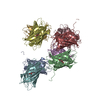
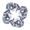
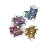
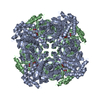
| Title | Crystal structure of particulate methane monooxygenase from Methylomicrobium alcaliphilum 20Z | |||||||||
 Components Components |
| |||||||||
 Keywords Keywords | OXIDOREDUCTASE / copper dependent methane monooxygenase | |||||||||
| Function / homology |  Function and homology information Function and homology informationmethane monooxygenase (soluble) / methane monooxygenase [NAD(P)H] activity / metal ion binding Similarity search - Function | |||||||||
| Biological species |  Methylomicrobium alcaliphilum 20Z (bacteria) Methylomicrobium alcaliphilum 20Z (bacteria) | |||||||||
| Method |  X-RAY DIFFRACTION / SYNCHROTRON / MOLECULAR REPLACEMENT / Resolution: 2.704 Å X-RAY DIFFRACTION / SYNCHROTRON / MOLECULAR REPLACEMENT / Resolution: 2.704 Å | |||||||||
 Authors Authors | Ro, S.Y. / Rosenzweig, A.C. | |||||||||
| Funding support |  United States, 2items United States, 2items
| |||||||||
 Citation Citation | Journal: J. Biol. Chem. / Year: 2018 Title: From micelles to bicelles: Effect of the membrane on particulate methane monooxygenase activity. Authors: Ro, S.Y. / Ross, M.O. / Deng, Y.W. / Batelu, S. / Lawton, T.J. / Hurley, J.D. / Stemmler, T.L. / Hoffman, B.M. / Rosenzweig, A.C. | |||||||||
| History |
|
- Structure visualization
| Structure viewer | Molecule: MolmilJmol/JSmol |
|---|
- Downloads & links
-Download
| PDBx/mmCIF format | 6cxh.cif.gz | 295 KB | Display | PDBx/mmCIF format |
|---|---|---|---|---|
| PDB format | pdb6cxh.ent.gz | 239.6 KB | Display | PDB format |
| PDBx/mmJSON format | 6cxh.json.gz | Tree view | PDBx/mmJSON format | |
| Others |  Other downloads Other downloads |
-Validation report
| Arichive directory | https://data.pdbj.org/pub/pdb/validation_reports/cx/6cxhftp://data.pdbj.org/pub/pdb/validation_reports/cx/6cxh | HTTPS FTP |
|---|
-Related structure data
| Related structure data | 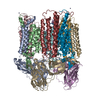 3rgbS S: Starting model for refinement |
|---|---|
| Similar structure data |


-Links
 PDBj
PDBj- Assembly
| Deposited unit | 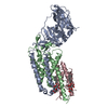
| ||||||||
|---|---|---|---|---|---|---|---|---|---|
| 1 | 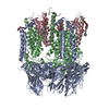
| ||||||||
| Unit cell |
|
-Components
| #1: Protein | Mass: 45602.125 Da / Num. of mol.: 1 / Source method: isolated from a natural source Source: (natural) Methylomicrobium alcaliphilum 20Z (bacteria)Strain: DSM 19304 / NCIMB 14124 / VKM B-2133 / 20Z References: UniProt: G4SZ64, methane monooxygenase (soluble) |
|---|---|
| #2: Protein | Mass: 28277.934 Da / Num. of mol.: 1 / Source method: isolated from a natural source Source: (natural) Methylomicrobium alcaliphilum 20Z (bacteria)Strain: DSM 19304 / NCIMB 14124 / VKM B-2133 / 20Z References: UniProt: G4SZ63, methane monooxygenase (soluble) |
| #3: Protein | Mass: 28874.301 Da / Num. of mol.: 1 / Source method: isolated from a natural source Source: (natural) Methylomicrobium alcaliphilum 20Z (bacteria)Strain: DSM 19304 / NCIMB 14124 / VKM B-2133 / 20Z References: UniProt: G4SZ62, methane monooxygenase (soluble) |
| #4: Chemical | ChemComp-CU /   Mass: 63.546 Da / Num. of mol.: 1 / Source method: obtained synthetically / Formula: Cu Mass: 63.546 Da / Num. of mol.: 1 / Source method: obtained synthetically / Formula: Cu |
| #5: Chemical |   Mass: 494.573 Da / Num. of mol.: 2 / Source method: obtained synthetically / Formula: C23H42O11 / Comment: detergent*YM Mass: 494.573 Da / Num. of mol.: 2 / Source method: obtained synthetically / Formula: C23H42O11 / Comment: detergent*YM |
-Experimental details
-Experiment
| Experiment | Method: X-RAY DIFFRACTION / Number of used crystals: 1 |
|---|
- Sample preparation
| Crystal | Density Matthews: 4.25 Å3/Da / Density % sol: 71.04 % |
|---|---|
| Crystal grow | Temperature: 293 K / Method: vapor diffusion, sitting drop / Details: 2.8 M AmSO4, 0.2 M MES, pH 6 |
-Data collection
| Diffraction | Mean temperature: 100 K | |||||||||
|---|---|---|---|---|---|---|---|---|---|---|
| Diffraction source | Source: SYNCHROTRON / Site: APS / Beamline: 21-ID-D / Wavelength: 1.033288, 1.377602 | |||||||||
| Detector | Type: DECTRIS EIGER X 9M / Detector: PIXEL / Date: Jul 28, 2016 | |||||||||
| Radiation | Protocol: SINGLE WAVELENGTH / Monochromatic (M) / Laue (L): M / Scattering type: x-ray | |||||||||
| Radiation wavelength |
| |||||||||
| Reflection | Resolution: 2.7→40 Å / Num. obs: 46989 / % possible obs: 99.1 % / Redundancy: 9 % / CC1/2: 0.999 / Rpim(I) all: 0.025 / Rrim(I) all: 0.07 / Net I/σ(I): 40.6 | |||||||||
| Reflection shell | Resolution: 2.7→2.8 Å / Redundancy: 6.4 % / Mean I/σ(I) obs: 11.2 / CC1/2: 0.984 / Rpim(I) all: 0.22 / Rrim(I) all: 0.588 / % possible all: 92 |
- Processing
| Software |
| ||||||||||||||||||||||||||||||||||||||||||||||||||||||||||||||||||||||||||||||||||||||||||||||||||
|---|---|---|---|---|---|---|---|---|---|---|---|---|---|---|---|---|---|---|---|---|---|---|---|---|---|---|---|---|---|---|---|---|---|---|---|---|---|---|---|---|---|---|---|---|---|---|---|---|---|---|---|---|---|---|---|---|---|---|---|---|---|---|---|---|---|---|---|---|---|---|---|---|---|---|---|---|---|---|---|---|---|---|---|---|---|---|---|---|---|---|---|---|---|---|---|---|---|---|---|
| Refinement | Method to determine structure: MOLECULAR REPLACEMENT Starting model: 3RGB Resolution: 2.704→39.579 Å / SU ML: 0.31 / Cross valid method: FREE R-VALUE / σ(F): 1.36 / Phase error: 29.27
| ||||||||||||||||||||||||||||||||||||||||||||||||||||||||||||||||||||||||||||||||||||||||||||||||||
| Solvent computation | Shrinkage radii: 0.9 Å / VDW probe radii: 1.11 Å | ||||||||||||||||||||||||||||||||||||||||||||||||||||||||||||||||||||||||||||||||||||||||||||||||||
| Refinement step | Cycle: LAST / Resolution: 2.704→39.579 Å
| ||||||||||||||||||||||||||||||||||||||||||||||||||||||||||||||||||||||||||||||||||||||||||||||||||
| Refine LS restraints |
| ||||||||||||||||||||||||||||||||||||||||||||||||||||||||||||||||||||||||||||||||||||||||||||||||||
| LS refinement shell |
| ||||||||||||||||||||||||||||||||||||||||||||||||||||||||||||||||||||||||||||||||||||||||||||||||||
| Refinement TLS params. | Method: refined / Origin x: -168.5951 Å / Origin y: -171.7598 Å / Origin z: 8.9381 Å
| ||||||||||||||||||||||||||||||||||||||||||||||||||||||||||||||||||||||||||||||||||||||||||||||||||
| Refinement TLS group | Selection details: all |
