Method to determine structure: SAD / Resolution: 2.8→29.46 Å / Cor.coef. Fo:Fc: 0.885 / Cor.coef. Fo:Fc free: 0.857 / SU B: 45.312 / SU ML: 0.357 / Cross valid method: THROUGHOUT / σ(F): 0 / ESU R: 0.608 / ESU R Free: 0.365 / Stereochemistry target values: MAXIMUM LIKELIHOOD Details: HYDROGENS HAVE BEEN ADDED IN THE RIDING POSITIONS U VALUES : RESIDUAL ONLY
Rfactor
Num. reflection
% reflection
Selection details
Rfree
0.29606
5932
5 %
RANDOM
Rwork
0.2686
-
-
-
obs
0.26997
112261
91.18 %
-
all
-
123120
-
-
Solvent computation
Ion probe radii: 0.8 Å / Shrinkage radii: 0.8 Å / VDW probe radii: 1.4 Å / Solvent model: MASK
Displacement parameters
Biso mean: 74.167 Å2
Baniso -1
Baniso -2
Baniso -3
1-
0.04 Å2
0 Å2
0 Å2
2-
-
0.04 Å2
0 Å2
3-
-
-
-0.08 Å2
Refinement step
Cycle: LAST / Resolution: 2.8→29.46 Å
Protein
Nucleic acid
Ligand
Solvent
Total
Num. atoms
19716
0
18
3
19737
Refine LS restraints
Refine-ID
Type
Dev ideal
Dev ideal target
Number
X-RAY DIFFRACTION
r_bond_refined_d
0.015
0.022
20379
X-RAY DIFFRACTION
r_bond_other_d
X-RAY DIFFRACTION
r_angle_refined_deg
1.621
1.93
27822
X-RAY DIFFRACTION
r_angle_other_deg
X-RAY DIFFRACTION
r_dihedral_angle_1_deg
7.803
5
2424
X-RAY DIFFRACTION
r_dihedral_angle_2_deg
33.594
22.375
897
X-RAY DIFFRACTION
r_dihedral_angle_3_deg
20.863
15
3090
X-RAY DIFFRACTION
r_dihedral_angle_4_deg
17.22
15
126
X-RAY DIFFRACTION
r_chiral_restr
0.122
0.2
3012
X-RAY DIFFRACTION
r_gen_planes_refined
0.007
0.021
15624
X-RAY DIFFRACTION
r_gen_planes_other
X-RAY DIFFRACTION
r_nbd_refined
X-RAY DIFFRACTION
r_nbd_other
X-RAY DIFFRACTION
r_nbtor_refined
X-RAY DIFFRACTION
r_nbtor_other
X-RAY DIFFRACTION
r_xyhbond_nbd_refined
X-RAY DIFFRACTION
r_xyhbond_nbd_other
X-RAY DIFFRACTION
r_metal_ion_refined
X-RAY DIFFRACTION
r_metal_ion_other
X-RAY DIFFRACTION
r_symmetry_vdw_refined
X-RAY DIFFRACTION
r_symmetry_vdw_other
X-RAY DIFFRACTION
r_symmetry_hbond_refined
X-RAY DIFFRACTION
r_symmetry_hbond_other
X-RAY DIFFRACTION
r_symmetry_metal_ion_refined
X-RAY DIFFRACTION
r_symmetry_metal_ion_other
X-RAY DIFFRACTION
r_mcbond_it
0.509
1.5
12132
X-RAY DIFFRACTION
r_mcbond_other
X-RAY DIFFRACTION
r_mcangle_it
0.998
2
19662
X-RAY DIFFRACTION
r_scbond_it
1.342
3
8247
X-RAY DIFFRACTION
r_scangle_it
2.296
4.5
8160
X-RAY DIFFRACTION
r_rigid_bond_restr
X-RAY DIFFRACTION
r_sphericity_free
X-RAY DIFFRACTION
r_sphericity_bonded
Refine LS restraints NCS
Refine-ID: X-RAY DIFFRACTION
Ens-ID
Dom-ID
Auth asym-ID
Number
Type
Rms dev position (Å)
Weight position
1
1
A
3018
tightpositional
0.06
0.05
1
2
I
3018
tightpositional
0.06
0.05
2
1
E
3018
tightpositional
0.06
0.05
2
2
I
3018
tightpositional
0.06
0.05
3
1
B
1797
tightpositional
0.05
0.05
3
2
J
1797
tightpositional
0.05
0.05
4
1
F
1797
tightpositional
0.05
0.05
4
2
J
1797
tightpositional
0.05
0.05
5
1
C
1760
tightpositional
0.04
0.05
5
2
K
1760
tightpositional
0.04
0.05
6
1
G
1565
tightpositional
0.04
0.05
6
2
K
1565
tightpositional
0.04
0.05
1
1
A
3018
tightthermal
0.11
0.5
1
2
I
3018
tightthermal
0.11
0.5
2
1
E
3018
tightthermal
0.11
0.5
2
2
I
3018
tightthermal
0.11
0.5
3
1
B
1797
tightthermal
0.09
0.5
3
2
J
1797
tightthermal
0.09
0.5
4
1
F
1797
tightthermal
0.08
0.5
4
2
J
1797
tightthermal
0.08
0.5
5
1
C
1760
tightthermal
0.06
0.5
5
2
K
1760
tightthermal
0.06
0.5
6
1
G
1565
tightthermal
0.05
0.5
6
2
K
1565
tightthermal
0.05
0.5
LS refinement shell
Resolution: 2.801→2.873 Å / Total num. of bins used: 20
Rfactor
Num. reflection
% reflection
Rfree
0.497
227
-
Rwork
0.501
4053
-
obs
-
-
45.47 %
Refinement TLS params.
Method: refined / Refine-ID: X-RAY DIFFRACTION
ID
L11 (°2)
L12 (°2)
L13 (°2)
L22 (°2)
L23 (°2)
L33 (°2)
S11 (Å °)
S12 (Å °)
S13 (Å °)
S21 (Å °)
S22 (Å °)
S23 (Å °)
S31 (Å °)
S32 (Å °)
S33 (Å °)
T11 (Å2)
T12 (Å2)
T13 (Å2)
T22 (Å2)
T23 (Å2)
T33 (Å2)
Origin x (Å)
Origin y (Å)
Origin z (Å)
1
0.9175
-0.6652
-0.1625
1.0311
-0.0271
0.4196
-0.0033
-0.054
-0.0723
0.0479
0.0249
0.0813
-0.0486
-0.0133
-0.0216
0.0675
0.0071
-0.0084
0.1037
0.0271
0.0178
34.93
125.1728
73.5053
2
3.6094
0.1305
0.2609
0.6059
0.0635
0.4553
-0.0218
0.3171
0.108
0.0554
0.069
-0.1533
0.1225
0.0319
-0.0472
0.0888
0.0861
-0.0116
0.1746
0.0459
0.133
62.4959
111.0155
64.8193
3
3.7524
-1.771
0.327
1.9277
0.1289
1.5938
-0.1084
-0.244
0.3919
0.2544
0.1246
-0.3103
-0.1007
-0.0745
-0.0162
0.0914
0.0373
-0.1152
0.1471
-0.0078
0.284
73.0971
127.3594
78.3243
4
1.4884
0.0176
0.8638
0.556
0.2493
1.0393
-0.0196
0.0453
0.1301
0.0158
-0.0141
-0.1493
-0.1176
-0.0108
0.0337
0.0729
-0.0448
-0.0341
0.0645
0.0338
0.065
32.6164
153.2931
41.6825
5
0.4889
-0.0419
-0.0112
0.6122
-0.3857
1.1734
0.0391
0.0263
-0.0688
-0.0383
0.0914
0.1042
0.3097
-0.082
-0.1304
0.1002
-0.0369
-0.0281
0.1481
0.0011
0.0903
30.4984
111.8331
33.5961
6
3.2644
-0.7908
0.3244
0.7545
-0.3
0.9152
-0.0484
-0.2708
0.1321
-0.08
-0.0796
-0.2536
0.2086
0.299
0.128
0.0846
0.0226
0.0726
0.3954
0.0149
0.1375
58.7001
122.5836
22.1735
7
3.5768
-0.5147
-0.9484
0.3416
0.0345
1.5412
-0.0549
0.2131
-0.1205
0.0035
-0.068
-0.2043
-0.0873
0.3207
0.1229
0.1174
-0.1055
-0.0852
0.1783
0.0703
0.3034
62.7471
153.8805
53.2468
8
4.17
0.0387
-1.1477
2.0386
-0.367
2.426
-0.0858
0.1105
-0.1864
-0.1081
-0.042
-0.2791
0.4121
0.116
0.1278
0.2082
0.2321
0.0536
0.4337
-0.0566
0.1646
67.3517
101.4316
28.5977
9
3.0222
0.8339
0.5676
1.8294
0.3179
2.699
-0.1333
0.2502
0.2012
-0.2119
0.0818
-0.1548
-0.0913
0.4103
0.0515
0.1248
-0.1731
0.0908
0.5254
0.1464
0.3891
69.3929
157.8828
30.8542
Refinement TLS group
ID
Refine-ID
Refine TLS-ID
Auth asym-ID
Auth seq-ID
1
X-RAY DIFFRACTION
1
A
33 - 414
2
X-RAY DIFFRACTION
2
B
7 - 245
3
X-RAY DIFFRACTION
3
C
45 - 286
4
X-RAY DIFFRACTION
3
B
664
5
X-RAY DIFFRACTION
4
E
33 - 414
6
X-RAY DIFFRACTION
5
I
33 - 414
7
X-RAY DIFFRACTION
6
J
7 - 245
8
X-RAY DIFFRACTION
7
F
7 - 245
9
X-RAY DIFFRACTION
8
K
45 - 286
10
X-RAY DIFFRACTION
8
J
664
11
X-RAY DIFFRACTION
9
G
45 - 286
12
X-RAY DIFFRACTION
9
F
664
+
About Yorodumi
-
News
-
Feb 9, 2022. New format data for meta-information of EMDB entries
New format data for meta-information of EMDB entries
Version 3 of the EMDB header file is now the official format.
The previous official version 1.9 will be removed from the archive.
In the structure databanks used in Yorodumi, some data are registered as the other names, "COVID-19 virus" and "2019-nCoV". Here are the details of the virus and the list of structure data.
Jan 31, 2019. EMDB accession codes are about to change! (news from PDBe EMDB page)
EMDB accession codes are about to change! (news from PDBe EMDB page)
The allocation of 4 digits for EMDB accession codes will soon come to an end. Whilst these codes will remain in use, new EMDB accession codes will include an additional digit and will expand incrementally as the available range of codes is exhausted. The current 4-digit format prefixed with “EMD-” (i.e. EMD-XXXX) will advance to a 5-digit format (i.e. EMD-XXXXX), and so on. It is currently estimated that the 4-digit codes will be depleted around Spring 2019, at which point the 5-digit format will come into force.
The EM Navigator/Yorodumi systems omit the EMD- prefix.
Related info.:Q: What is EMD? / ID/Accession-code notation in Yorodumi/EM Navigator
Yorodumi is a browser for structure data from EMDB, PDB, SASBDB, etc.
This page is also the successor to EM Navigator detail page, and also detail information page/front-end page for Omokage search.
The word "yorodu" (or yorozu) is an old Japanese word meaning "ten thousand". "mi" (miru) is to see.
Related info.:EMDB / PDB / SASBDB / Comparison of 3 databanks / Yorodumi Search / Aug 31, 2016. New EM Navigator & Yorodumi / Yorodumi Papers / Jmol/JSmol / Function and homology information / Changes in new EM Navigator and Yorodumi
 Movie
Movie Controller

 Yorodumi
 Open data
 Basic information
 Components
 Keywords
 Function and homology information
 Methylococcus capsulatus (bacteria)
 X-RAY DIFFRACTION /
  Authors
 Citation
 Structure visualization
 Downloads & links
 Other downloads
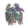


 PDBj
PDBj
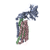
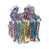
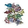
 Assembly


 Mass: 63.546 Da / Num. of mol.: 3 / Source method: obtained synthetically / Formula: Cu
 Mass: 65.409 Da / Num. of mol.: 9 / Source method: obtained synthetically / Formula: Zn
 Mass: 127.092 Da / Num. of mol.: 3 / Source method: obtained synthetically / Formula: Cu2
 Sample preparation
 / Beamline: 5.0.1 / Wavelength: 1.377,1.280
 Processing