 Movie
Movie Controller

[English] 日本語
 Yorodumi
- PDB-4pi0: Crystal structure of particulate methane monooxygenase from Methy... -
+ Open data

- Basic information
| Entry | Database: PDB / ID: 4pi0 | ||||||
|---|---|---|---|---|---|---|---|
| Title | Crystal structure of particulate methane monooxygenase from Methylocystis sp. ATCC 49242 (Rockwell) soaked in copper | ||||||
 Components Components |
| ||||||
 Keywords Keywords | OXIDOREDUCTASE / Bacterial Proteins / Binding Sites / Copper / Zinc / Methylocystaceae / Oxygenases / Protein Binding | ||||||
| Function / homology |  Function and homology information Function and homology informationHelix hairpin bin / Particulate methane monooxygenase subunit c2. Chain: C / particulate methane monooxygenase, chain B / Ammonia/particulate methane monooxygenase, subunit A / Particulate methane monooxygenase, b subunit. Chain: A, domain 3 / Particulate methane monooxygenase, b subunit. Chain: A, domain 1 / Glutathione S-transferase Yfyf (Class Pi); Chain A, domain 2 / Helix Hairpins / Jelly Rolls / Up-down Bundle ...Helix hairpin bin / Particulate methane monooxygenase subunit c2. Chain: C / particulate methane monooxygenase, chain B / Ammonia/particulate methane monooxygenase, subunit A / Particulate methane monooxygenase, b subunit. Chain: A, domain 3 / Particulate methane monooxygenase, b subunit. Chain: A, domain 1 / Glutathione S-transferase Yfyf (Class Pi); Chain A, domain 2 / Helix Hairpins / Jelly Rolls / Up-down Bundle / Immunoglobulin-like / Sandwich / Orthogonal Bundle / Mainly Beta / Mainly Alpha Similarity search - Domain/homology | ||||||
| Biological species |  Methylocystis sp. ATCC 49242 (bacteria) Methylocystis sp. ATCC 49242 (bacteria) | ||||||
| Method |  X-RAY DIFFRACTION / SYNCHROTRON / MOLECULAR REPLACEMENT / Resolution: 3.15 Å X-RAY DIFFRACTION / SYNCHROTRON / MOLECULAR REPLACEMENT / Resolution: 3.15 Å | ||||||
 Authors Authors | Sirajuddin, S. / Rosenzweig, A.C. | ||||||
| Funding support |  United States, 1items United States, 1items
| ||||||
 Citation Citation | Journal: J.Biol.Chem. / Year: 2014 Title: Effects of zinc on particulate methane monooxygenase activity and structure. Authors: Sirajuddin, S. / Barupala, D. / Helling, S. / Marcus, K. / Stemmler, T.L. / Rosenzweig, A.C. | ||||||
| History |
|
- Structure visualization
| Structure viewer | Molecule: MolmilJmol/JSmol |
|---|
- Downloads & links
-Download
| PDBx/mmCIF format | 4pi0.cif.gz | 513.1 KB | Display | PDBx/mmCIF format |
|---|---|---|---|---|
| PDB format | pdb4pi0.ent.gz | 420.1 KB | Display | PDB format |
| PDBx/mmJSON format | 4pi0.json.gz | Tree view | PDBx/mmJSON format | |
| Others |  Other downloads Other downloads |
-Validation report
| Arichive directory | https://data.pdbj.org/pub/pdb/validation_reports/pi/4pi0ftp://data.pdbj.org/pub/pdb/validation_reports/pi/4pi0 | HTTPS FTP |
|---|
-Related structure data
| Related structure data | 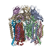 4phzC  4pi2C 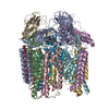 3rfrS C: citing same article ( S: Starting model for refinement |
|---|---|
| Similar structure data |


-Links
 PDBj
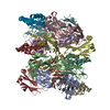
PDBj- Assembly
| Deposited unit | 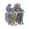
| |||||||||||||||||||||||||||||||||||||||||||||||||||||||||||||||||||||||||||||||||||||||||||||||||||||||||||||||||||||||||||||||||||||||||||||||||||||||||||||||||||||||||||||||||||||||||||||||
|---|---|---|---|---|---|---|---|---|---|---|---|---|---|---|---|---|---|---|---|---|---|---|---|---|---|---|---|---|---|---|---|---|---|---|---|---|---|---|---|---|---|---|---|---|---|---|---|---|---|---|---|---|---|---|---|---|---|---|---|---|---|---|---|---|---|---|---|---|---|---|---|---|---|---|---|---|---|---|---|---|---|---|---|---|---|---|---|---|---|---|---|---|---|---|---|---|---|---|---|---|---|---|---|---|---|---|---|---|---|---|---|---|---|---|---|---|---|---|---|---|---|---|---|---|---|---|---|---|---|---|---|---|---|---|---|---|---|---|---|---|---|---|---|---|---|---|---|---|---|---|---|---|---|---|---|---|---|---|---|---|---|---|---|---|---|---|---|---|---|---|---|---|---|---|---|---|---|---|---|---|---|---|---|---|---|---|---|---|---|---|---|---|
| 1 |
| |||||||||||||||||||||||||||||||||||||||||||||||||||||||||||||||||||||||||||||||||||||||||||||||||||||||||||||||||||||||||||||||||||||||||||||||||||||||||||||||||||||||||||||||||||||||||||||||
| Unit cell |
| |||||||||||||||||||||||||||||||||||||||||||||||||||||||||||||||||||||||||||||||||||||||||||||||||||||||||||||||||||||||||||||||||||||||||||||||||||||||||||||||||||||||||||||||||||||||||||||||
| Noncrystallographic symmetry (NCS) | NCS domain:
NCS domain segments: Refine code: 1
|