 Movie
Movie Controller


+ Open data

- Basic information
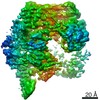
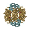
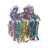
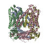
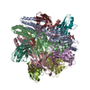
| Entry | Database: PDB / ID: 7k78 | ||||||
|---|---|---|---|---|---|---|---|
| Title | antibody and nucleosome complex | ||||||
 Components Components |
| ||||||
 Keywords Keywords | DNA BINDING PROTEIN/DNA / nucleosome / DNA BINDING PROTEIN / DNA BINDING PROTEIN-DNA complex | ||||||
| Function / homology |  Function and homology information Function and homology informationHATs acetylate histones / RNA polymerase I upstream activating factor complex / Condensation of Prophase Chromosomes / : / : / : / Assembly of the ORC complex at the origin of replication / HDACs deacetylate histones / Oxidative Stress Induced Senescence / Recruitment and ATM-mediated phosphorylation of repair and signaling proteins at DNA double strand breaks ...HATs acetylate histones / RNA polymerase I upstream activating factor complex / Condensation of Prophase Chromosomes / : / : / : / Assembly of the ORC complex at the origin of replication / HDACs deacetylate histones / Oxidative Stress Induced Senescence / Recruitment and ATM-mediated phosphorylation of repair and signaling proteins at DNA double strand breaks / RMTs methylate histone arginines / DNA damage tolerance / SUMOylation of chromatin organization proteins / RNA Polymerase I Promoter Escape / positive regulation of transcription by RNA polymerase I / nucleolar large rRNA transcription by RNA polymerase I / Estrogen-dependent gene expression / Ub-specific processing proteases / structural constituent of chromatin / nucleosome / heterochromatin formation / nucleosome assembly / chromatin organization / protein heterodimerization activity / DNA repair / regulation of DNA-templated transcription / negative regulation of transcription by RNA polymerase II / DNA binding / identical protein binding / nucleus Similarity search - Function | ||||||
| Biological species |   | ||||||
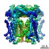
| Method | ELECTRON MICROSCOPY / single particle reconstruction / cryo EM / Resolution: 3.1 Å | ||||||
 Authors Authors | Ruifang, G. / Yawen, B. | ||||||
 Citation Citation | Journal: Nat Commun / Year: 2021 Title: Structural and dynamic mechanisms of CBF3-guided centromeric nucleosome formation. Authors: Ruifang Guan / Tengfei Lian / Bing-Rui Zhou / Emily He / Carl Wu / Martin Singleton / Yawen Bai /   Abstract: Accurate chromosome segregation relies on the specific centromeric nucleosome-kinetochore interface. In budding yeast, the centromere CBF3 complex guides the deposition of CENP-A, an H3 variant, to ...Accurate chromosome segregation relies on the specific centromeric nucleosome-kinetochore interface. In budding yeast, the centromere CBF3 complex guides the deposition of CENP-A, an H3 variant, to form the centromeric nucleosome in a DNA sequence-dependent manner. Here, we determine the structures of the centromeric nucleosome containing the native CEN3 DNA and the CBF3core bound to the canonical nucleosome containing an engineered CEN3 DNA. The centromeric nucleosome core structure contains 115 base pair DNA including a CCG motif. The CBF3core specifically recognizes the nucleosomal CCG motif through the Gal4 domain while allosterically altering the DNA conformation. Cryo-EM, modeling, and mutational studies reveal that the CBF3core forms dynamic interactions with core histones H2B and CENP-A in the CEN3 nucleosome. Our results provide insights into the structure of the budding yeast centromeric nucleosome and the mechanism of its assembly, which have implications for analogous processes of human centromeric nucleosome formation. | ||||||
| History |
|
- Structure visualization
| Movie |
Movie viewer |
|---|---|
| Structure viewer | Molecule: MolmilJmol/JSmol |
- Downloads & links
-Download
| PDBx/mmCIF format | 7k78.cif.gz | 325.7 KB | Display | PDBx/mmCIF format |
|---|---|---|---|---|
| PDB format | pdb7k78.ent.gz | 250.9 KB | Display | PDB format |
| PDBx/mmJSON format | 7k78.json.gz | Tree view | PDBx/mmJSON format | |
| Others |  Other downloads Other downloads |
-Validation report
| Arichive directory | https://data.pdbj.org/pub/pdb/validation_reports/k7/7k78ftp://data.pdbj.org/pub/pdb/validation_reports/k7/7k78 | HTTPS FTP |
|---|
-Related structure data
| Related structure data | 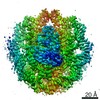 22696MC 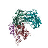 7k79C  7k7gC M: map data used to model this data C: citing same article ( |
|---|---|
| Similar structure data |


-Links
 PDBj
PDBj


- Assembly
| Deposited unit | 
|
|---|---|
| 1 |
|
-Components
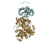
-Protein , 4 types, 8 molecules AEBFCGDH
| #1: Protein | Mass: 15791.588 Da / Num. of mol.: 2 Source method: isolated from a genetically manipulated source Source: (gene. exp.) Production host:  Escherichia phage EcSzw-2 (virus) Escherichia phage EcSzw-2 (virus)#2: Protein | Mass: 11395.390 Da / Num. of mol.: 2 Source method: isolated from a genetically manipulated source Source: (gene. exp.) Strain: ATCC 204508 / S288c / Gene: HHF1, YBR009C, YBR0122, HHF2, YNL030W, N2752 / Production host: Escherichia phage EcSzw-2 (virus) / References: UniProt: P02309#3: Protein | Mass: 14013.177 Da / Num. of mol.: 2 Source method: isolated from a genetically manipulated source Source: (gene. exp.) Strain: ATCC 204508 / S288c / Gene: HTA1, H2A1, SPT11, YDR225W, YD9934.10 / Production host: Escherichia phage EcSzw-2 (virus) / References: UniProt: P04911#4: Protein | Mass: 14280.362 Da / Num. of mol.: 2 Source method: isolated from a genetically manipulated source Source: (gene. exp.) Strain: ATCC 204508 / S288c / Gene: HTB1, H2B1, SPT12, YDR224C, YD9934.09C / Production host: Escherichia phage EcSzw-2 (virus) / References: UniProt: P02293 |
|---|
-DNA chain , 2 types, 2 molecules IJ
| #5: DNA chain | Mass: 42219.285 Da / Num. of mol.: 1 Source method: isolated from a genetically manipulated source Source: (gene. exp.) Production host: Escherichia phage EcSzw-2 (virus) |
|---|---|
| #6: DNA chain | Mass: 41679.848 Da / Num. of mol.: 1 Source method: isolated from a genetically manipulated source Source: (gene. exp.) Production host: Escherichia phage EcSzw-2 (virus) |
-Antibody , 1 types, 2 molecules KL
| #7: Antibody | Mass: 29030.146 Da / Num. of mol.: 2 Source method: isolated from a genetically manipulated source Source: (gene. exp.) Escherichia phage EcSzw-2 (virus) |
|---|
-Details
| Has protein modification | Y |
|---|
-Experimental details
-Experiment
| Experiment | Method: ELECTRON MICROSCOPY |
|---|---|
| EM experiment | Aggregation state: PARTICLE / 3D reconstruction method: single particle reconstruction |
- Sample preparation
| Component | Name: scFv and nucleosome complex / Type: COMPLEX / Entity ID: all / Source: NATURAL |
|---|---|
| Source (natural) | Organism: |
| Buffer solution | pH: 7.3 |
| Specimen | Embedding applied: NO / Shadowing applied: NO / Staining applied: NO / Vitrification applied: YES |
| Vitrification | Cryogen name: NITROGEN / Humidity: 100 % |
- Electron microscopy imaging
| Experimental equipment |  Model: Titan Krios / Image courtesy: FEI Company |
|---|---|
| Microscopy | Model: FEI TITAN KRIOS |
| Electron gun | Electron source:  FIELD EMISSION GUN / Accelerating voltage: 300 kV / Illumination mode: SPOT SCAN FIELD EMISSION GUN / Accelerating voltage: 300 kV / Illumination mode: SPOT SCAN |
| Electron lens | Mode: BRIGHT FIELD |
| Image recording | Electron dose: 71 e/Å2 / Film or detector model: GATAN K2 SUMMIT (4k x 4k) |
- Processing
| CTF correction | Type: NONE |
|---|---|
| 3D reconstruction | Resolution: 3.1 Å / Resolution method: FSC 0.143 CUT-OFF / Num. of particles: 143164 / Symmetry type: POINT |
