 Movie
Movie Controller

[English] 日本語
 Yorodumi
- PDB-3chx: Crystal structure of Methylosinus trichosporium OB3b particulate ... -
+ Open data

- Basic information
| Entry | Database: PDB / ID: 3chx | ||||||
|---|---|---|---|---|---|---|---|
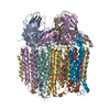
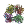
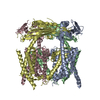
| Title | Crystal structure of Methylosinus trichosporium OB3b particulate methane monooxygenase (pMMO) | ||||||
 Components Components |
| ||||||
 Keywords Keywords | MEMBRANE PROTEIN / METHANE / BETA BARREL | ||||||
| Function / homology |  Function and homology information Function and homology information | ||||||
| Biological species |  Methylosinus trichosporium (bacteria) Methylosinus trichosporium (bacteria) | ||||||
| Method |  X-RAY DIFFRACTION / SYNCHROTRON / MOLECULAR REPLACEMENT / molecular replacement / Resolution: 3.9 Å X-RAY DIFFRACTION / SYNCHROTRON / MOLECULAR REPLACEMENT / molecular replacement / Resolution: 3.9 Å | ||||||
 Authors Authors | Hakemian, A.S. | ||||||
 Citation Citation | Journal: Biochemistry / Year: 2008 Title: The metal centers of particulate methane monooxygenase from Methylosinus trichosporium OB3b. Authors: Hakemian, A.S. / Kondapalli, K.C. / Telser, J. / Hoffman, B.M. / Stemmler, T.L. / Rosenzweig, A.C. | ||||||
| History |
|
- Structure visualization
| Structure viewer | Molecule: MolmilJmol/JSmol |
|---|
- Downloads & links
-Download
| PDBx/mmCIF format | 3chx.cif.gz | 426.9 KB | Display | PDBx/mmCIF format |
|---|---|---|---|---|
| PDB format | pdb3chx.ent.gz | 325 KB | Display | PDB format |
| PDBx/mmJSON format | 3chx.json.gz | Tree view | PDBx/mmJSON format | |
| Others |  Other downloads Other downloads |
-Validation report
| Arichive directory | https://data.pdbj.org/pub/pdb/validation_reports/ch/3chxftp://data.pdbj.org/pub/pdb/validation_reports/ch/3chx | HTTPS FTP |
|---|
-Related structure data
| Related structure data | 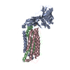 1yewS S: Starting model for refinement |
|---|---|
| Similar structure data |


-Links
 PDBj
PDBj- Assembly
| Deposited unit | 
| ||||||||||||
|---|---|---|---|---|---|---|---|---|---|---|---|---|---|
| 1 |
| ||||||||||||
| Unit cell |
| ||||||||||||
| Noncrystallographic symmetry (NCS) | NCS domain: (Details: chain AAAA,CCCC, using strict) NCS oper:
|
-Components
-Protein , 3 types, 9 molecules AEIBFJCGK
| #1: Protein | Mass: 43181.145 Da / Num. of mol.: 3 Source method: isolated from a genetically manipulated source Source: (gene. exp.) Methylosinus trichosporium (bacteria) / Gene: pmoB / References: UniProt: Q9KX50#2: Protein | Mass: 28524.338 Da / Num. of mol.: 3 Source method: isolated from a genetically manipulated source Source: (gene. exp.) Methylosinus trichosporium (bacteria) / Gene: pmoA / References: UniProt: Q50541#3: Protein | Mass: 29024.742 Da / Num. of mol.: 3 Source method: isolated from a genetically manipulated source Source: (gene. exp.) Methylosinus trichosporium (bacteria) / Gene: pmoC / References: UniProt: Q9KX51 |
|---|
-Protein/peptide , 2 types, 6 molecules DHLMNO
| #4: Protein/peptide | Mass: 1720.111 Da / Num. of mol.: 3 Source method: isolated from a genetically manipulated source Source: (gene. exp.) Methylosinus trichosporium (bacteria)#5: Protein/peptide | Mass: 2230.741 Da / Num. of mol.: 3 Source method: isolated from a genetically manipulated source Source: (gene. exp.) Methylosinus trichosporium (bacteria) |
|---|
-Non-polymers , 1 types, 9 molecules 
| #6: Chemical | ChemComp-CU /  Mass: 63.546 Da / Num. of mol.: 9 / Source method: obtained synthetically / Formula: Cu Mass: 63.546 Da / Num. of mol.: 9 / Source method: obtained synthetically / Formula: Cu |
|---|
-Details
| Sequence details | AUTHORS STATE THAT IT WAS NOT POSSIBLE TO ASSIGN PEPTIDES IN CHAINS D,H,L.M,N, AND O TO A ...AUTHORS STATE THAT IT WAS NOT POSSIBLE TO ASSIGN PEPTIDES IN CHAINS D,H,L.M,N, AND O TO A PARTICULAR |
|---|
-Experimental details
-Experiment
| Experiment | Method: X-RAY DIFFRACTION / Number of used crystals: 1 |
|---|
- Sample preparation
| Crystal | Density Matthews: 3.4 Å3/Da / Density % sol: 63.83 % |
|---|---|
| Crystal grow | Temperature: 293 K / Method: vapor diffusion, sitting drop / pH: 6.5 Details: 0.1 M cacodylate, 10% PEG3000, 0.25 M manganese chloride, pH 6.5, VAPOR DIFFUSION, SITTING DROP, temperature 293K |
-Data collection
| Diffraction | Mean temperature: 100 K |
|---|---|
| Diffraction source | Source: SYNCHROTRON / Site: APS  / Beamline: 23-ID-B / Wavelength: 0.9794 Å / Beamline: 23-ID-B / Wavelength: 0.9794 Å |
| Detector | Type: MARMOSAIC 300 mm CCD / Detector: CCD / Date: Dec 20, 2005 |
| Radiation | Monochromator: double crystal monochromator and K-B pair of biomorph mirrors for vertical and horizontal focusing Protocol: SINGLE WAVELENGTH / Monochromatic (M) / Laue (L): M / Scattering type: x-ray |
| Radiation wavelength | Wavelength: 0.9794 Å / Relative weight: 1 |
| Reflection | Resolution: 3.9→37.969 Å / Num. obs: 39753 / % possible obs: 99.8 % / Observed criterion σ(F): 0 / Observed criterion σ(I): 0 / Redundancy: 7.4 % / Rmerge(I) obs: 0.086 / Rsym value: 0.086 / Net I/σ(I): 7.5 |
| Reflection shell | Resolution: 3.9→4.11 Å / Redundancy: 7.5 % / Rmerge(I) obs: 0.376 / Mean I/σ(I) obs: 2 / Num. measured all: 43098 / Num. unique all: 5723 / Rsym value: 0.376 / % possible all: 100 |
-Phasing
| Phasing | Method: molecular replacement | |||||||||
|---|---|---|---|---|---|---|---|---|---|---|
| Phasing MR | Model details: Phaser MODE: MR_AUTO
|
- Processing
| Software |
| ||||||||||||||||||||||||||||
|---|---|---|---|---|---|---|---|---|---|---|---|---|---|---|---|---|---|---|---|---|---|---|---|---|---|---|---|---|---|
| Refinement | Method to determine structure: MOLECULAR REPLACEMENT Starting model: PDB ENTRY 1YEW Resolution: 3.9→38 Å / FOM work R set: 0.623 / Isotropic thermal model: anisotropic / Cross valid method: THROUGHOUT / σ(F): 0 / σ(I): 0 / Stereochemistry target values: Engh & Huber
| ||||||||||||||||||||||||||||
| Solvent computation | Bsol: 114.657 Å2 | ||||||||||||||||||||||||||||
| Displacement parameters | Biso mean: 141.814 Å2
| ||||||||||||||||||||||||||||
| Refinement step | Cycle: LAST / Resolution: 3.9→38 Å
| ||||||||||||||||||||||||||||
| Refine LS restraints |
| ||||||||||||||||||||||||||||
| Refine LS restraints NCS | Rms: 0 / Type: strict / Weight: 300 | ||||||||||||||||||||||||||||
| LS refinement shell | Resolution: 3.9→4.04 Å
| ||||||||||||||||||||||||||||
| Xplor file |
|
