 Movie
Movie Controller


+ Open data

- Basic information
| Entry | Database: PDB / ID: 1jjo | ||||||
|---|---|---|---|---|---|---|---|
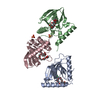
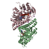
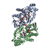
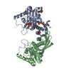
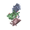
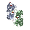
| Title | Crystal Structure of Mouse Neuroserpin (Cleaved form) | ||||||
 Components Components | (NEUROSERPIN) x 3 | ||||||
 Keywords Keywords | SIGNALING PROTEIN / Serpin / Serine protease inhibitor / Neuronal serpin | ||||||
| Function / homology |  Function and homology information Function and homology informationcytoplasmic vesicle lumen / serine-type endopeptidase inhibitor activity / positive regulation of neuron projection development / secretory granule lumen / perikaryon / neuronal cell body / : Similarity search - Function | ||||||
| Biological species |  | ||||||
| Method |  X-RAY DIFFRACTION / MOLECULAR REPLACEMENT / Resolution: 3.06 Å X-RAY DIFFRACTION / MOLECULAR REPLACEMENT / Resolution: 3.06 Å | ||||||
 Authors Authors | Briand, C. / Kozlov, S.V. / Sonderegger, P. / Gruetter, M.G. | ||||||
 Citation Citation | Journal: FEBS Lett. / Year: 2001 Title: Crystal structure of neuroserpin: a neuronal serpin involved in a conformational disease. Authors: Briand, C. / Kozlov, S.V. / Sonderegger, P. / Grutter, M.G. | ||||||
| History |
|
- Structure visualization
| Structure viewer | Molecule: MolmilJmol/JSmol |
|---|
- Downloads & links
-Download
| PDBx/mmCIF format | 1jjo.cif.gz | 134 KB | Display | PDBx/mmCIF format |
|---|---|---|---|---|
| PDB format | pdb1jjo.ent.gz | 106.3 KB | Display | PDB format |
| PDBx/mmJSON format | 1jjo.json.gz | Tree view | PDBx/mmJSON format | |
| Others |  Other downloads Other downloads |
-Validation report
| Arichive directory | https://data.pdbj.org/pub/pdb/validation_reports/jj/1jjoftp://data.pdbj.org/pub/pdb/validation_reports/jj/1jjo | HTTPS FTP |
|---|
-Related structure data
| Similar structure data |
|---|


-Links
 PDBj
PDBj
- Assembly
| Deposited unit | 
| ||||||||
|---|---|---|---|---|---|---|---|---|---|
| 1 |
| ||||||||
| Unit cell |
|
-Components
| #1: Protein/peptide | Mass: 4416.040 Da / Num. of mol.: 2 Source method: isolated from a genetically manipulated source Source: (gene. exp.)  #2: Protein | Mass: 29630.404 Da / Num. of mol.: 2 Source method: isolated from a genetically manipulated source Source: (gene. exp.) #3: Protein/peptide | Mass: 3969.749 Da / Num. of mol.: 2 Source method: isolated from a genetically manipulated source Source: (gene. exp.) |
|---|
-Experimental details
-Experiment
| Experiment | Method: X-RAY DIFFRACTION / Number of used crystals: 1 |
|---|
- Sample preparation
| Crystal | Density Matthews: 2.07 Å3/Da / Density % sol: 40.63 % | |||||||||||||||||||||||||||||||||||
|---|---|---|---|---|---|---|---|---|---|---|---|---|---|---|---|---|---|---|---|---|---|---|---|---|---|---|---|---|---|---|---|---|---|---|---|---|
| Crystal grow | Temperature: 293 K / Method: vapor diffusion, sitting drop / pH: 6.1 Details: 25% PEG8000, 0.2M H2PO4(NH4), 0.1M Tris-HCl, pH 6.1, VAPOR DIFFUSION, SITTING DROP, temperature 293K | |||||||||||||||||||||||||||||||||||
| Crystal grow | *PLUS Temperature: 20 ℃ | |||||||||||||||||||||||||||||||||||
| Components of the solutions | *PLUS
|
-Data collection
| Diffraction | Mean temperature: 100 K |
|---|---|
| Diffraction source | Source: ROTATING ANODE / Type: ENRAF-NONIUS FR571 / Wavelength: 1.5418 Å |
| Detector | Type: MARRESEARCH / Detector: IMAGE PLATE / Date: Jul 6, 1999 |
| Radiation | Monochromator: mirror, Prophysics XRM-216 / Protocol: SINGLE WAVELENGTH / Monochromatic (M) / Laue (L): M / Scattering type: x-ray |
| Radiation wavelength | Wavelength: 1.5418 Å / Relative weight: 1 |
| Reflection | Resolution: 3.06→16.86 Å / Num. all: 11124 / Num. obs: 9419 / % possible obs: 79.7 % / Observed criterion σ(F): 0 / Observed criterion σ(I): 2 / Redundancy: 2.57 % / Rsym value: 0.155 / Net I/σ(I): 7 |
| Reflection shell | Resolution: 3.06→3.1 Å / Redundancy: 2.3 % / Mean I/σ(I) obs: 2 / Num. unique all: 417 / Rsym value: 0.421 / % possible all: 94.6 |
| Reflection | *PLUS Lowest resolution: 17 Å / Rmerge(I) obs: 0.155 |
| Reflection shell | *PLUS % possible obs: 74.6 % / Num. unique obs: 7975 / Rmerge(I) obs: 0.422 / Mean I/σ(I) obs: 2 |
- Processing
| Software |
| |||||||||||||||||||||||||
|---|---|---|---|---|---|---|---|---|---|---|---|---|---|---|---|---|---|---|---|---|---|---|---|---|---|---|
| Refinement | Method to determine structure: MOLECULAR REPLACEMENT Starting model: homology model based on all available cleaved serpin structures Resolution: 3.06→16.89 Å / Isotropic thermal model: anisotropic / σ(F): 0 / Stereochemistry target values: Engh & Huber / Details: Bulk solvent correction, strict NCS
| |||||||||||||||||||||||||
| Refinement step | Cycle: LAST / Resolution: 3.06→16.89 Å
| |||||||||||||||||||||||||
| Refine LS restraints |
| |||||||||||||||||||||||||
| Software | *PLUS Name: CNS / Version: 0.9 / Classification: refinement | |||||||||||||||||||||||||
| Refinement | *PLUS σ(F): 0 / % reflection Rfree: 4.3 % / Rfactor obs: 0.232 | |||||||||||||||||||||||||
| Solvent computation | *PLUS | |||||||||||||||||||||||||
| Displacement parameters | *PLUS |
