 Movie
Movie Controller

[English] 日本語
 Yorodumi
- PDB-2o8m: Crystal structure of the S139A mutant of Hepatitis C Virus NS3/4A... -
+ Open data

- Basic information
| Entry | Database: PDB / ID: 2o8m | ||||||
|---|---|---|---|---|---|---|---|
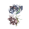
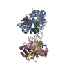
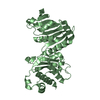
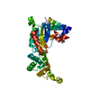
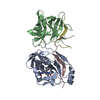
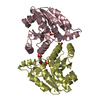
| Title | Crystal structure of the S139A mutant of Hepatitis C Virus NS3/4A protease | ||||||
 Components Components | (Protease) x 2 | ||||||
 Keywords Keywords | VIRAL PROTEIN / SERINE PROTEASE / NS3 / NS4A / HEPATITIS C VIRUS | ||||||
| Function / homology |  Function and homology information Function and homology informationsymbiont-mediated suppression of host JAK-STAT cascade via inhibition of STAT activity / positive regulation of hexokinase activity / positive regulation of metabolic process / translocation of peptides or proteins into host cell cytoplasm / symbiont-mediated perturbation of host cellular process / regulation of primary metabolic process / Toll-like receptor 2 binding / viral capsid assembly / hepacivirin / adhesion receptor-mediated virion attachment to host cell ...symbiont-mediated suppression of host JAK-STAT cascade via inhibition of STAT activity / positive regulation of hexokinase activity / positive regulation of metabolic process / translocation of peptides or proteins into host cell cytoplasm / symbiont-mediated perturbation of host cellular process / regulation of primary metabolic process / Toll-like receptor 2 binding / viral capsid assembly / hepacivirin / adhesion receptor-mediated virion attachment to host cell / TBC/RABGAPs / host cell mitochondrial membrane / host cell lipid droplet / symbiont-mediated transformation of host cell / symbiont-mediated suppression of host TRAF-mediated signal transduction / positive regulation of cytokinesis / symbiont-mediated perturbation of host cell cycle G1/S transition checkpoint / negative regulation of protein secretion / symbiont-mediated suppression of host JAK-STAT cascade via inhibition of STAT1 activity / endoplasmic reticulum-Golgi intermediate compartment membrane / symbiont-mediated suppression of host cytoplasmic pattern recognition receptor signaling pathway via inhibition of MAVS activity / SH3 domain binding / kinase binding / ribonucleoside triphosphate phosphatase activity / nucleoside-triphosphate phosphatase / viral nucleocapsid / channel activity / monoatomic ion transmembrane transport / clathrin-dependent endocytosis of virus by host cell / entry receptor-mediated virion attachment to host cell / Hydrolases; Acting on peptide bonds (peptidases); Cysteine endopeptidases / RNA helicase activity / host cell perinuclear region of cytoplasm / host cell endoplasmic reticulum membrane / RNA helicase / symbiont-mediated suppression of host type I interferon-mediated signaling pathway / ribonucleoprotein complex / serine-type endopeptidase activity / viral translational frameshifting / symbiont-mediated activation of host autophagy / RNA-directed RNA polymerase / cysteine-type endopeptidase activity / viral RNA genome replication / RNA-directed RNA polymerase activity / fusion of virus membrane with host endosome membrane / viral envelope / virion attachment to host cell / host cell nucleus / host cell plasma membrane / virion membrane / structural molecule activity / negative regulation of transcription by RNA polymerase II / ATP hydrolysis activity / proteolysis / RNA binding / zinc ion binding / ATP binding Similarity search - Function | ||||||
| Biological species |  Hepatitis C virus Hepatitis C virus | ||||||
| Method |  X-RAY DIFFRACTION / FOURIER SYNTHESIS / Resolution: 2 Å X-RAY DIFFRACTION / FOURIER SYNTHESIS / Resolution: 2 Å | ||||||
 Authors Authors | Fischmann, T.O. / Prongay, A.J. / Madison, V.M. / Yao, N. | ||||||
 Citation Citation | Journal: J.Med.Chem. / Year: 2007 Title: Discovery of the HCV NS3/4A protease inhibitor (1R,5S)-N-[3-amino-1-(cyclobutylmethyl)-2,3-dioxopropyl]-3- [2(S)-[[[(1,1-dimethylethyl)amino]carbonyl]amino]-3,3-dimethyl-1-oxobutyl]- 6,6- ...Title: Discovery of the HCV NS3/4A protease inhibitor (1R,5S)-N-[3-amino-1-(cyclobutylmethyl)-2,3-dioxopropyl]-3- [2(S)-[[[(1,1-dimethylethyl)amino]carbonyl]amino]-3,3-dimethyl-1-oxobutyl]- 6,6-dimethyl-3-azabicyclo[3.1.0]hexan-2(S)-carboxamide (Sch 503034) II. Key steps in structure-based optimization Authors: Prongay, A.J. / Guo, Z. / Yao, N. / Pichardo, J. / Fischmann, T. / Strickland, C. / Myers, J. / Weber, P.C. / Beyer, B.M. / Ingram, R. / Hong, Z. / Prosise, W.W. / Ramanathan, L. / Taremi, S. ...Authors: Prongay, A.J. / Guo, Z. / Yao, N. / Pichardo, J. / Fischmann, T. / Strickland, C. / Myers, J. / Weber, P.C. / Beyer, B.M. / Ingram, R. / Hong, Z. / Prosise, W.W. / Ramanathan, L. / Taremi, S.S. / Yarosh-Tomaine, T. / Zhang, R. / Senior, M. / Yang, R.S. / Malcolm, B. / Arasappan, A. / Bennett, F. / Bogen, S.L. / Chen, K. / Jao, E. / Liu, Y.T. / Lovey, R.G. / Saksena, A.K. / Venkatraman, S. / Girijavallabhan, V. / Njoroge, F.G. / Madison, V. | ||||||
| History |
|
- Structure visualization
| Structure viewer | Molecule: MolmilJmol/JSmol |
|---|
- Downloads & links
-Download
| PDBx/mmCIF format | 2o8m.cif.gz | 90.1 KB | Display | PDBx/mmCIF format |
|---|---|---|---|---|
| PDB format | pdb2o8m.ent.gz | 67.3 KB | Display | PDB format |
| PDBx/mmJSON format | 2o8m.json.gz | Tree view | PDBx/mmJSON format | |
| Others |  Other downloads Other downloads |
-Validation report
| Arichive directory | https://data.pdbj.org/pub/pdb/validation_reports/o8/2o8mftp://data.pdbj.org/pub/pdb/validation_reports/o8/2o8m | HTTPS FTP |
|---|
-Related structure data
| Related structure data |  2oboC 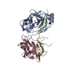 2obqC 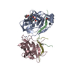 2oc0C 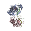 2oc1C 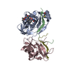 2oc7C 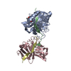 2oc8C C: citing same article ( |
|---|---|
| Similar structure data |


-Links
 PDBj
PDBj

- Assembly
| Deposited unit | 
| ||||||||
|---|---|---|---|---|---|---|---|---|---|
| 1 |
| ||||||||
| Unit cell |
|
-Components
| #1: Protein | Mass: 21217.225 Da / Num. of mol.: 2 / Mutation: S149A Source method: isolated from a genetically manipulated source Source: (gene. exp.) Hepatitis C virus / Genus: Hepacivirus / Gene: HCV / Production host:  References: UniProt: Q9ELS8, UniProt: P27958*PLUS, Hydrolases; Acting on peptide bonds (peptidases); Cysteine endopeptidases #2: Protein/peptide | Mass: 2410.104 Da / Num. of mol.: 2 / Source method: obtained synthetically / References: UniProt: P27958 #3: Chemical |   Mass: 65.409 Da / Num. of mol.: 2 / Source method: obtained synthetically / Formula: Zn Mass: 65.409 Da / Num. of mol.: 2 / Source method: obtained synthetically / Formula: Zn#4: Chemical |   Mass: 22.990 Da / Num. of mol.: 2 / Source method: obtained synthetically / Formula: Na Mass: 22.990 Da / Num. of mol.: 2 / Source method: obtained synthetically / Formula: Na#5: Water | ChemComp-HOH / |  Mass: 18.015 Da / Num. of mol.: 235 / Source method: isolated from a natural source / Formula: H2O Mass: 18.015 Da / Num. of mol.: 235 / Source method: isolated from a natural source / Formula: H2O |
|---|
-Experimental details
-Experiment
| Experiment | Method: X-RAY DIFFRACTION / Number of used crystals: 1 |
|---|
- Sample preparation
| Crystal | Density Matthews: 3.87 Å3/Da / Density % sol: 68.21 % |
|---|---|
| Crystal grow | Temperature: 277 K / Method: vapor diffusion, hanging drop Details: 1.25 to 1.5M NaCl, 0.1 M MES, .1M Na/K PO4, 5mM beta-mercaptoethanol, pH 5.6-6.2, VAPOR DIFFUSION, HANGING DROP, temperature 277K |
-Data collection
| Diffraction | Mean temperature: 200 K |
|---|---|
| Diffraction source | Source: ROTATING ANODE / Type: RIGAKU RU200 / Wavelength: 1.5418 Å |
| Detector | Type: RIGAKU RAXIS IIC / Detector: IMAGE PLATE / Date: Aug 1, 1997 / Details: Osmic mirrors |
| Radiation | Monochromator: Osmic mirrors / Protocol: SINGLE WAVELENGTH / Monochromatic (M) / Laue (L): M / Scattering type: x-ray |
| Radiation wavelength | Wavelength: 1.5418 Å / Relative weight: 1 |
| Reflection | Resolution: 2→50 Å / Num. obs: 46014 / % possible obs: 94.1 % / Observed criterion σ(F): -3 / Observed criterion σ(I): -1 / Redundancy: 3 % / Rmerge(I) obs: 0.042 / Rsym value: 0.0489 / Χ2: 0.887 / Net I/σ(I): 19.4 |
| Reflection shell | Resolution: 2→2.03 Å / Redundancy: 1.4 % / Rmerge(I) obs: 0.572 / Mean I/σ(I) obs: 1.1 / Num. unique all: 1429 / Χ2: 1.015 / % possible all: 58.7 |
- Processing
| Software |
| ||||||||||||||||||||||||||||
|---|---|---|---|---|---|---|---|---|---|---|---|---|---|---|---|---|---|---|---|---|---|---|---|---|---|---|---|---|---|
| Refinement | Method to determine structure: FOURIER SYNTHESIS / Resolution: 2→50 Å / Isotropic thermal model: Isotropic / Cross valid method: THROUGHOUT / σ(F): -1 / σ(I): -1 / Stereochemistry target values: Engh & Huber
| ||||||||||||||||||||||||||||
| Displacement parameters | Biso mean: 39.674 Å2 | ||||||||||||||||||||||||||||
| Refinement step | Cycle: LAST / Resolution: 2→50 Å
| ||||||||||||||||||||||||||||
| LS refinement shell | Resolution: 2→2 Å / Rfactor Rfree: 0.2688 / Rfactor Rwork: 0.2356 |
