- PDB-2o88: Crystal structure of the N114A mutant of ABL-SH3 domain complexed... -
+
Open data
ID or keywords:
Loading...
-
Basic information
Entry
Database: PDB / ID: 2o88
Title
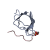
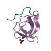
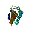
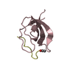
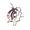
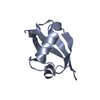
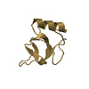
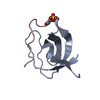
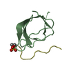
Crystal structure of the N114A mutant of ABL-SH3 domain complexed with a designed high-affinity peptide ligand: implications for SH3-ligand interactions
Components
P41 peptide
Proto-oncogene tyrosine-protein kinase ABL1
Keywords
SIGNALING PROTEIN / SH3 domain High affinity peptide complex
Function / homology
Function and homology information
protein localization to cytoplasmic microtubule plus-end / DNA conformation change / response to epinephrine / phospholipase C-inhibiting G protein-coupled receptor signaling pathway / negative regulation of ubiquitin-protein transferase activity / podocyte apoptotic process / regulation of postsynaptic specialization assembly / positive regulation of phospholipase C/protein kinase C signal transduction / regulation of modification of synaptic structure / nicotinate-nucleotide adenylyltransferase activity ...protein localization to cytoplasmic microtubule plus-end / DNA conformation change / response to epinephrine / phospholipase C-inhibiting G protein-coupled receptor signaling pathway / negative regulation of ubiquitin-protein transferase activity / podocyte apoptotic process / regulation of postsynaptic specialization assembly / positive regulation of phospholipase C/protein kinase C signal transduction / regulation of modification of synaptic structure / nicotinate-nucleotide adenylyltransferase activity / delta-catenin binding / mitochondrial depolarization / Role of ABL in ROBO-SLIT signaling / positive regulation of extracellular matrix organization / neuropilin signaling pathway / neuropilin binding / regulation of cell motility / bubble DNA binding / positive regulation of establishment of T cell polarity / cellular response to dopamine / positive regulation of blood vessel branching / proline-rich region binding / positive regulation of dendrite development / mitogen-activated protein kinase binding / regulation of Cdc42 protein signal transduction / regulation of hematopoietic stem cell differentiation / syntaxin binding / regulation of axon extension / regulation of T cell differentiation / positive regulation of cell migration involved in sprouting angiogenesis / Myogenesis / HDR through Single Strand Annealing (SSA) / platelet-derived growth factor receptor-beta signaling pathway / RUNX2 regulates osteoblast differentiation / Fc-gamma receptor signaling pathway involved in phagocytosis / vascular endothelial cell response to oscillatory fluid shear stress / myoblast proliferation / regulation of endocytosis / negative regulation of long-term synaptic potentiation / regulation of microtubule polymerization / associative learning / cardiac muscle cell proliferation / positive regulation of focal adhesion assembly / actin monomer binding / ephrin receptor signaling pathway / cellular response to transforming growth factor beta stimulus / positive regulation of vasoconstriction / regulation of cell adhesion / endothelial cell migration / RHO GTPases Activate WASPs and WAVEs / negative regulation of double-strand break repair via homologous recombination / positive regulation of T cell migration / mismatch repair / ephrin receptor binding / positive regulation of stress fiber assembly / four-way junction DNA binding / ruffle / signal transduction in response to DNA damage / phosphotyrosine residue binding / positive regulation of substrate adhesion-dependent cell spreading / actin filament polymerization / positive regulation of endothelial cell migration / integrin-mediated signaling pathway / SH2 domain binding / response to endoplasmic reticulum stress / Turbulent (oscillatory, disturbed) flow shear stress activates signaling by PIEZO1 and integrins in endothelial cells / protein kinase C binding / protein modification process / regulation of autophagy / protein serine/threonine kinase activator activity / regulation of actin cytoskeleton organization / non-specific protein-tyrosine kinase / FCGR3A-mediated phagocytosis / non-membrane spanning protein tyrosine kinase activity / Regulation of actin dynamics for phagocytic cup formation / intrinsic apoptotic signaling pathway in response to DNA damage / positive regulation of fibroblast proliferation / cellular response to hydrogen peroxide / epidermal growth factor receptor signaling pathway / enzyme activator activity / autophagy / sequence-specific double-stranded DNA binding / kinase activity / Cyclin D associated events in G1 / actin filament binding / actin cytoskeleton / positive regulation of neuron apoptotic process / manganese ion binding / mitotic cell cycle / Recruitment and ATM-mediated phosphorylation of repair and signaling proteins at DNA double strand breaks / positive regulation of cytosolic calcium ion concentration / Factors involved in megakaryocyte development and platelet production / MLL4 and MLL3 complexes regulate expression of PPARG target genes in adipogenesis and hepatic steatosis / growth cone / nuclear membrane / RUNX1 regulates transcription of genes involved in differentiation of HSCs / cellular response to oxidative stress / actin cytoskeleton organization / protein tyrosine kinase activity / response to oxidative stress Similarity search - Function
In the structure databanks used in Yorodumi, some data are registered as the other names, "COVID-19 virus" and "2019-nCoV". Here are the details of the virus and the list of structure data.
Jan 31, 2019. EMDB accession codes are about to change! (news from PDBe EMDB page)
EMDB accession codes are about to change! (news from PDBe EMDB page)
The allocation of 4 digits for EMDB accession codes will soon come to an end. Whilst these codes will remain in use, new EMDB accession codes will include an additional digit and will expand incrementally as the available range of codes is exhausted. The current 4-digit format prefixed with “EMD-” (i.e. EMD-XXXX) will advance to a 5-digit format (i.e. EMD-XXXXX), and so on. It is currently estimated that the 4-digit codes will be depleted around Spring 2019, at which point the 5-digit format will come into force.
The EM Navigator/Yorodumi systems omit the EMD- prefix.
Related info.:Q: What is EMD? / ID/Accession-code notation in Yorodumi/EM Navigator
Yorodumi is a browser for structure data from EMDB, PDB, SASBDB, etc.
This page is also the successor to EM Navigator detail page, and also detail information page/front-end page for Omokage search.
The word "yorodu" (or yorozu) is an old Japanese word meaning "ten thousand". "mi" (miru) is to see.
Related info.:EMDB / PDB / SASBDB / Comparison of 3 databanks / Yorodumi Search / Aug 31, 2016. New EM Navigator & Yorodumi / Yorodumi Papers / Jmol/JSmol / Function and homology information / Changes in new EM Navigator and Yorodumi
 Movie
Movie Controller

 Yorodumi
 Open data
 Basic information
 Components
 Keywords
 Function and homology information
 Homo sapiens (human)
 X-RAY DIFFRACTION /
  Authors
 Citation
 Structure visualization
 Downloads & links
 Other downloads
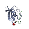
 PDBj
PDBj


 Assembly


 Mass: 96.063 Da / Num. of mol.: 2 / Source method: obtained synthetically / Formula: SO4
 Mass: 18.015 Da / Num. of mol.: 67 / Source method: isolated from a natural source / Formula: H2O
 Sample preparation
 Processing