 Movie
Movie Controller


+ Open data

- Basic information
| Entry | Database: PDB / ID: 6u97 | ||||||
|---|---|---|---|---|---|---|---|
| Title | Structure of OmcF_H47I mutant | ||||||
 Components Components | Lipoprotein cytochrome c, 1 heme-binding site | ||||||
 Keywords Keywords | ELECTRON TRANSPORT / OmcF / redox-Bohr effect | ||||||
| Function / homology |  Function and homology information Function and homology informationplasma membrane-derived thylakoid lumen / electron transfer activity / iron ion binding / heme binding Similarity search - Function | ||||||
| Biological species |  Geobacter sulfurreducens (bacteria) Geobacter sulfurreducens (bacteria) | ||||||
| Method |  X-RAY DIFFRACTION / SYNCHROTRON / MOLECULAR REPLACEMENT / Resolution: 1.13 Å X-RAY DIFFRACTION / SYNCHROTRON / MOLECULAR REPLACEMENT / Resolution: 1.13 Å | ||||||
 Authors Authors | Pokkuluri, P.R. | ||||||
| Funding support |  United States, 1items United States, 1items
| ||||||
 Citation Citation | Journal: Front Microbiol / Year: 2019 Title: Modulation of the Redox Potential and Electron/Proton Transfer Mechanisms in the Outer Membrane Cytochrome OmcF FromGeobacter sulfurreducens. Authors: Teixeira, L.R. / Cordas, C.M. / Fonseca, M.P. / Duke, N.E.C. / Pokkuluri, P.R. / Salgueiro, C.A. | ||||||
| History |
|
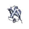
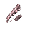
- Structure visualization
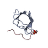
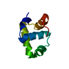
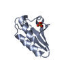
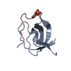
| Structure viewer | Molecule: MolmilJmol/JSmol |
|---|
- Downloads & links
-Download
| PDBx/mmCIF format | 6u97.cif.gz | 74.2 KB | Display | PDBx/mmCIF format |
|---|---|---|---|---|
| PDB format | pdb6u97.ent.gz | 45.7 KB | Display | PDB format |
| PDBx/mmJSON format | 6u97.json.gz | Tree view | PDBx/mmJSON format | |
| Others |  Other downloads Other downloads |
-Validation report
| Arichive directory | https://data.pdbj.org/pub/pdb/validation_reports/u9/6u97ftp://data.pdbj.org/pub/pdb/validation_reports/u9/6u97 | HTTPS FTP |
|---|
-Related structure data
| Similar structure data |
|---|


-Links
 PDBj
PDBj


- Assembly
| Deposited unit | 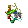
| ||||||||||||
|---|---|---|---|---|---|---|---|---|---|---|---|---|---|
| 1 |
| ||||||||||||
| Unit cell |
|
-Components
| #1: Protein | Mass: 8427.596 Da / Num. of mol.: 1 / Mutation: H47I Source method: isolated from a genetically manipulated source Source: (gene. exp.) Geobacter sulfurreducens (strain ATCC 51573 / DSM 12127 / PCA) (bacteria)Strain: ATCC 51573 / DSM 12127 / PCA / Gene: omcF, GSU2432 / Production host: |
|---|---|
| #2: Chemical | ChemComp-SO4 /   Mass: 96.063 Da / Num. of mol.: 1 / Source method: obtained synthetically / Formula: SO4 Mass: 96.063 Da / Num. of mol.: 1 / Source method: obtained synthetically / Formula: SO4 |
| #3: Chemical | ChemComp-HEM /   Mass: 616.487 Da / Num. of mol.: 1 / Source method: obtained synthetically / Formula: C34H32FeN4O4 / Feature type: SUBJECT OF INVESTIGATION Mass: 616.487 Da / Num. of mol.: 1 / Source method: obtained synthetically / Formula: C34H32FeN4O4 / Feature type: SUBJECT OF INVESTIGATION |
| #4: Water | ChemComp-HOH /  Mass: 18.015 Da / Num. of mol.: 92 / Source method: isolated from a natural source / Formula: H2O Mass: 18.015 Da / Num. of mol.: 92 / Source method: isolated from a natural source / Formula: H2O |
| Has ligand of interest | Y |
| Has protein modification | Y |
-Experimental details
-Experiment
| Experiment | Method: X-RAY DIFFRACTION / Number of used crystals: 1 |
|---|
- Sample preparation
| Crystal | Density Matthews: 2.03 Å3/Da / Density % sol: 39.44 % / Description: needle clusters |
|---|---|
| Crystal grow | Temperature: 298 K / Method: vapor diffusion, hanging drop Details: 1.8 M ammonium sulfate, 0.1 M Hepes pH 7.5, 2% (v/v) PEG400 |
-Data collection
| Diffraction | Mean temperature: 100 K / Serial crystal experiment: N |
|---|---|
| Diffraction source | Source: SYNCHROTRON / Site: APS / Beamline: 22-ID / Wavelength: 0.9184 Å |
| Detector | Type: DECTRIS PILATUS3 S 6M / Detector: PIXEL / Date: Nov 20, 2018 |
| Radiation | Protocol: SINGLE WAVELENGTH / Monochromatic (M) / Laue (L): M / Scattering type: x-ray |
| Radiation wavelength | Wavelength: 0.9184 Å / Relative weight: 1 |
| Reflection | Resolution: 1→50 Å / Num. obs: 31673 / % possible obs: 99 % / Redundancy: 11 % / Biso Wilson estimate: 10.02 Å2 / CC1/2: 0.994 / Rmerge(I) obs: 0.054 / Rpim(I) all: 0.016 / Rrim(I) all: 0.056 / Χ2: 0.929 / Net I/σ(I): 42.5 |
| Reflection shell | Resolution: 1.13→1.15 Å / Redundancy: 6 % / Rmerge(I) obs: 0.304 / Mean I/σ(I) obs: 4.8 / Num. unique obs: 1901 / CC1/2: 0.963 / Rpim(I) all: 0.114 / Rrim(I) all: 0.327 / Χ2: 0.958 / % possible all: 95 |
- Processing
| Software |
| |||||||||||||||||||||||||||||||||||||||||||||||||
|---|---|---|---|---|---|---|---|---|---|---|---|---|---|---|---|---|---|---|---|---|---|---|---|---|---|---|---|---|---|---|---|---|---|---|---|---|---|---|---|---|---|---|---|---|---|---|---|---|---|---|
| Refinement | Method to determine structure: MOLECULAR REPLACEMENT / Resolution: 1.13→23.87 Å / SU ML: 0.0605 / Cross valid method: THROUGHOUT / σ(F): 1.42 / Phase error: 19.3043
| |||||||||||||||||||||||||||||||||||||||||||||||||
| Solvent computation | Shrinkage radii: 0.9 Å / VDW probe radii: 1.11 Å | |||||||||||||||||||||||||||||||||||||||||||||||||
| Displacement parameters | Biso mean: 13.97 Å2 | |||||||||||||||||||||||||||||||||||||||||||||||||
| Refinement step | Cycle: LAST / Resolution: 1.13→23.87 Å
| |||||||||||||||||||||||||||||||||||||||||||||||||
| Refine LS restraints |
| |||||||||||||||||||||||||||||||||||||||||||||||||
| LS refinement shell |
|
