- PDB-2nyg: Crystal structure of YokD protein from Bacillus subtilis -
+
Open data
ID or keywords:
Loading...
-
Basic information
Entry
Database: PDB / ID: 2nyg
Title
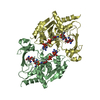
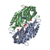
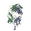
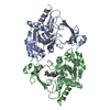
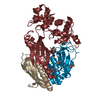
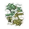
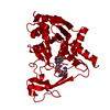
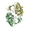
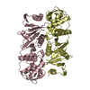
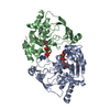
Crystal structure of YokD protein from Bacillus subtilis
Components
YokD protein
Keywords
STRUCTURAL GENOMICS / UNKNOWN FUNCTION / pfam02522 / NYSGXRC / 10116c / Aminoglycoside 3-N-acetyltransferase / PSI-2 / Protein Structure Initiative / New York SGX Research Center for Structural Genomics
Function / homology
Aminoglycoside N(3)-acetyltransferase / Aminoglycoside 3-N-acetyltransferase / Aminoglycoside 3-N-acetyltransferase-like / N-acetyltransferase activity / Transferases; Acyltransferases; Transferring groups other than aminoacyl groups / response to antibiotic / COENZYME A / SPbeta prophage-derived aminoglycoside N(3')-acetyltransferase-like protein YokD
Function and homology information
Biological species
Bacillus subtilis (bacteria)
Method
X-RAY DIFFRACTION / SYNCHROTRON / SAD / Resolution: 2.6 Å
Method to determine structure: SAD / Resolution: 2.6→49.04 Å / Rfactor Rfree error: 0.009 / Data cutoff high absF: 2587132.44 / Data cutoff low absF: 0 / Isotropic thermal model: RESTRAINED / Cross valid method: THROUGHOUT / σ(F): 0 / Stereochemistry target values: Engh & Huber Details: Residues listed as missing in Remark 465 are due to poor or lack of electron density. Residues with missing atoms listed in Remark 470 were modeled as ALA because of poor electron density.
In the structure databanks used in Yorodumi, some data are registered as the other names, "COVID-19 virus" and "2019-nCoV". Here are the details of the virus and the list of structure data.
Jan 31, 2019. EMDB accession codes are about to change! (news from PDBe EMDB page)
EMDB accession codes are about to change! (news from PDBe EMDB page)
The allocation of 4 digits for EMDB accession codes will soon come to an end. Whilst these codes will remain in use, new EMDB accession codes will include an additional digit and will expand incrementally as the available range of codes is exhausted. The current 4-digit format prefixed with “EMD-” (i.e. EMD-XXXX) will advance to a 5-digit format (i.e. EMD-XXXXX), and so on. It is currently estimated that the 4-digit codes will be depleted around Spring 2019, at which point the 5-digit format will come into force.
The EM Navigator/Yorodumi systems omit the EMD- prefix.
Related info.:Q: What is EMD? / ID/Accession-code notation in Yorodumi/EM Navigator
Yorodumi is a browser for structure data from EMDB, PDB, SASBDB, etc.
This page is also the successor to EM Navigator detail page, and also detail information page/front-end page for Omokage search.
The word "yorodu" (or yorozu) is an old Japanese word meaning "ten thousand". "mi" (miru) is to see.
Related info.:EMDB / PDB / SASBDB / Comparison of 3 databanks / Yorodumi Search / Aug 31, 2016. New EM Navigator & Yorodumi / Yorodumi Papers / Jmol/JSmol / Function and homology information / Changes in new EM Navigator and Yorodumi
 Movie
Movie Controller


 Open data
 Basic information
 Components
 Keywords
 Function and homology information
 X-RAY DIFFRACTION /
  Authors
 Citation
 Structure visualization
 Downloads & links
 Other downloads
 PDBj
PDBj


 Assembly
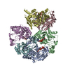


 Mass: 767.534 Da / Num. of mol.: 5 / Source method: obtained synthetically / Formula: C21H36N7O16P3S
 Mass: 18.015 Da / Num. of mol.: 223 / Source method: isolated from a natural source / Formula: H2O
 Sample preparation
 Processing