 Movie
Movie Controller

[English] 日本語
 Yorodumi
- PDB-3sma: A new N-acetyltransferase fold in the structure and mechanism of ... -
+ Open data

- Basic information
| Entry | Database: PDB / ID: 3sma | ||||||
|---|---|---|---|---|---|---|---|
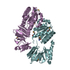
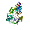
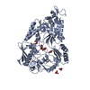
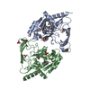
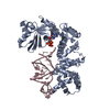
| Title | A new N-acetyltransferase fold in the structure and mechanism of the phosphonate biosynthetic enzyme FrbF | ||||||
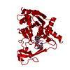
 Components Components | FrbF | ||||||
 Keywords Keywords | TRANSFERASE / N-acetyl transferase / acetyl CoA binding | ||||||
| Function / homology | Aminoglycoside N(3)-acetyltransferase / Aminoglycoside 3-N-acetyltransferase / Aminoglycoside 3-N-acetyltransferase-like / N-acetyltransferase activity / antibiotic biosynthetic process / Transferases; Acyltransferases; Transferring groups other than aminoacyl groups / response to antibiotic / ACETYL COENZYME *A / CMP-5'-(N-hydroxy-3-aminopropyl)phosphonate acetyltransferase Function and homology information Function and homology information | ||||||
| Biological species |  Streptomyces rubellomurinus (bacteria) Streptomyces rubellomurinus (bacteria) | ||||||
| Method |  X-RAY DIFFRACTION / SYNCHROTRON / MOLECULAR REPLACEMENT / Resolution: 2 Å X-RAY DIFFRACTION / SYNCHROTRON / MOLECULAR REPLACEMENT / Resolution: 2 Å | ||||||
 Authors Authors | Bae, B. / Nair, S.K. | ||||||
 Citation Citation | Journal: J.Biol.Chem. / Year: 2011 Title: New N-Acetyltransferase Fold in the Structure and Mechanism of the Phosphonate Biosynthetic Enzyme FrbF. Authors: Bae, B. / Cobb, R.E. / Desieno, M.A. / Zhao, H. / Nair, S.K. | ||||||
| History |
|
- Structure visualization
| Structure viewer | Molecule: MolmilJmol/JSmol |
|---|
- Downloads & links
-Download
| PDBx/mmCIF format | 3sma.cif.gz | 223.3 KB | Display | PDBx/mmCIF format |
|---|---|---|---|---|
| PDB format | pdb3sma.ent.gz | 180.6 KB | Display | PDB format |
| PDBx/mmJSON format | 3sma.json.gz | Tree view | PDBx/mmJSON format | |
| Others |  Other downloads Other downloads |
-Validation report
| Arichive directory | https://data.pdbj.org/pub/pdb/validation_reports/sm/3smaftp://data.pdbj.org/pub/pdb/validation_reports/sm/3sma | HTTPS FTP |
|---|
-Related structure data
| Similar structure data |
|---|
-Links
 PDBj
PDBj- Assembly
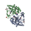
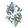
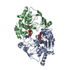
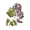
| Deposited unit | 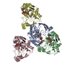
| ||||||||
|---|---|---|---|---|---|---|---|---|---|
| 1 | 
| ||||||||
| 2 | 
| ||||||||
| Unit cell |
|
-Components
| #1: Protein | Mass: 31586.535 Da / Num. of mol.: 4 Source method: isolated from a genetically manipulated source Source: (gene. exp.) Streptomyces rubellomurinus (bacteria) / Gene: frbF / Production host: #2: Chemical | ChemComp-ACO /   Mass: 809.571 Da / Num. of mol.: 4 / Source method: obtained synthetically / Formula: C23H38N7O17P3S Mass: 809.571 Da / Num. of mol.: 4 / Source method: obtained synthetically / Formula: C23H38N7O17P3S#3: Water | ChemComp-HOH / |  Mass: 18.015 Da / Num. of mol.: 511 / Source method: isolated from a natural source / Formula: H2O Mass: 18.015 Da / Num. of mol.: 511 / Source method: isolated from a natural source / Formula: H2O |
|---|
-Experimental details
-Experiment
| Experiment | Method: X-RAY DIFFRACTION / Number of used crystals: 1 |
|---|
- Sample preparation
| Crystal | Density Matthews: 2.19 Å3/Da / Density % sol: 43.95 % |
|---|---|
| Crystal grow | Temperature: 291 K / Method: vapor diffusion, hanging drop / pH: 7.7 Details: 0.2M magnesium chloride, 0.1 Tris, 20%-25% peg 4000, 20% glycerol, pH 7.7, VAPOR DIFFUSION, HANGING DROP, temperature 291K |
-Data collection
| Diffraction source | Source: SYNCHROTRON / Site: APS  / Beamline: 21-ID-D / Beamline: 21-ID-D |
|---|---|
| Detector | Type: MARMOSAIC 300 mm CCD / Detector: CCD / Date: Jun 12, 2009 |
| Radiation | Protocol: SINGLE WAVELENGTH / Monochromatic (M) / Laue (L): M / Scattering type: x-ray |
| Radiation wavelength | Relative weight: 1 |
| Reflection | Resolution: 2→45.34 Å / Num. all: 93253 / Num. obs: 74767 / Biso Wilson estimate: 26.44 Å2 |
- Processing
| Software |
| ||||||||||||||||||||||||||||||||||||||||||||||||||||||||||||||||||||||||||||||||||||||||||||||||||||||||||||||||||
|---|---|---|---|---|---|---|---|---|---|---|---|---|---|---|---|---|---|---|---|---|---|---|---|---|---|---|---|---|---|---|---|---|---|---|---|---|---|---|---|---|---|---|---|---|---|---|---|---|---|---|---|---|---|---|---|---|---|---|---|---|---|---|---|---|---|---|---|---|---|---|---|---|---|---|---|---|---|---|---|---|---|---|---|---|---|---|---|---|---|---|---|---|---|---|---|---|---|---|---|---|---|---|---|---|---|---|---|---|---|---|---|---|---|---|---|
| Refinement | Method to determine structure: MOLECULAR REPLACEMENT / Resolution: 2→45.34 Å / Cor.coef. Fo:Fc: 0.8846 / Cor.coef. Fo:Fc free: 0.8466 / Cross valid method: THROUGHOUT / σ(F): 0
| ||||||||||||||||||||||||||||||||||||||||||||||||||||||||||||||||||||||||||||||||||||||||||||||||||||||||||||||||||
| Displacement parameters | Biso mean: 35.93 Å2
| ||||||||||||||||||||||||||||||||||||||||||||||||||||||||||||||||||||||||||||||||||||||||||||||||||||||||||||||||||
| Refine analyze | Luzzati coordinate error obs: 0.254 Å | ||||||||||||||||||||||||||||||||||||||||||||||||||||||||||||||||||||||||||||||||||||||||||||||||||||||||||||||||||
| Refinement step | Cycle: LAST / Resolution: 2→45.34 Å
| ||||||||||||||||||||||||||||||||||||||||||||||||||||||||||||||||||||||||||||||||||||||||||||||||||||||||||||||||||
| Refine LS restraints |
| ||||||||||||||||||||||||||||||||||||||||||||||||||||||||||||||||||||||||||||||||||||||||||||||||||||||||||||||||||
| LS refinement shell | Resolution: 2→2.05 Å / Total num. of bins used: 20
|
