SEQUENCE THE CLOSEST SEQUENCE MATCH FOR THIS ENTRY IS AN UNIPROT ENTRY A6Y463_VIBCH, OF THE VIBRIO ... SEQUENCE THE CLOSEST SEQUENCE MATCH FOR THIS ENTRY IS AN UNIPROT ENTRY A6Y463_VIBCH, OF THE VIBRIO CHOLERAE RC385 STRAIN. AUTHORS STATE THAT THE SOURCE OF THEIR PROTEIN IS VIBRIO CHOLERAE O1 BIOVAR ELTOR STRAIN N16961, NCBI ACCESSION AAF94064, GI:9655358.
Resolution: 1.53→1.58 Å / Redundancy: 3.9 % / Mean I/σ(I) obs: 2.4 / Num. unique all: 12259 / Rsym value: 0.359 / % possible all: 76.5
-
Processing
Software
Name
Version
Classification
REFMAC
5.2.0019
refinement
SBC-Collect
datacollection
HKL-3000
datacollection
HKL-3000
datareduction
HKL-3000
datascaling
HKL-3000
phasing
SHELX
phasing
MLPHARE
phasing
RESOLVE
phasing
ARP/wARP
modelbuilding
Refinement
Method to determine structure: SAD / Resolution: 1.53→26.16 Å / Cor.coef. Fo:Fc: 0.975 / Cor.coef. Fo:Fc free: 0.96 / SU B: 2.773 / SU ML: 0.047 / Cross valid method: THROUGHOUT / σ(F): 0 / ESU R: 0.106 / ESU R Free: 0.079 Stereochemistry target values: MAXIMUM LIKELIHOOD WITH PHASES Details: Program PHENIX has also been used in refinement
Rfactor
Num. reflection
% reflection
Selection details
Rfree
0.183
7527
5 %
RANDOM
Rwork
0.143
-
-
-
all
0.145
142270
-
-
obs
0.145
142270
93.89 %
-
Solvent computation
Ion probe radii: 0.8 Å / Shrinkage radii: 0.8 Å / VDW probe radii: 1.4 Å / Solvent model: MASK
Displacement parameters
Biso mean: 21.199 Å2
Baniso -1
Baniso -2
Baniso -3
1-
-0.62 Å2
-0.18 Å2
-0.29 Å2
2-
-
0.89 Å2
1.12 Å2
3-
-
-
0.57 Å2
Refinement step
Cycle: LAST / Resolution: 1.53→26.16 Å
Protein
Nucleic acid
Ligand
Solvent
Total
Num. atoms
8197
0
94
1177
9468
Refine LS restraints
Refine-ID
Type
Dev ideal
Dev ideal target
Number
X-RAY DIFFRACTION
r_bond_refined_d
0.015
0.021
9589
X-RAY DIFFRACTION
r_bond_other_d
X-RAY DIFFRACTION
r_angle_refined_deg
1.578
1.948
13200
X-RAY DIFFRACTION
r_angle_other_deg
X-RAY DIFFRACTION
r_dihedral_angle_1_deg
6.13
5
1238
X-RAY DIFFRACTION
r_dihedral_angle_2_deg
40.448
24.981
526
X-RAY DIFFRACTION
r_dihedral_angle_3_deg
13.391
15
1604
X-RAY DIFFRACTION
r_dihedral_angle_4_deg
11.071
15
65
X-RAY DIFFRACTION
r_chiral_restr
0.112
0.2
1377
X-RAY DIFFRACTION
r_gen_planes_refined
0.007
0.02
7849
X-RAY DIFFRACTION
r_gen_planes_other
X-RAY DIFFRACTION
r_nbd_refined
0.233
0.2
4890
X-RAY DIFFRACTION
r_nbd_other
X-RAY DIFFRACTION
r_nbtor_refined
0.307
0.2
6690
X-RAY DIFFRACTION
r_nbtor_other
X-RAY DIFFRACTION
r_xyhbond_nbd_refined
0.178
0.2
1166
X-RAY DIFFRACTION
r_xyhbond_nbd_other
X-RAY DIFFRACTION
r_metal_ion_refined
0.124
0.2
2
X-RAY DIFFRACTION
r_metal_ion_other
X-RAY DIFFRACTION
r_symmetry_vdw_refined
0.226
0.2
102
X-RAY DIFFRACTION
r_symmetry_vdw_other
X-RAY DIFFRACTION
r_symmetry_hbond_refined
0.161
0.2
50
X-RAY DIFFRACTION
r_symmetry_hbond_other
X-RAY DIFFRACTION
r_symmetry_metal_ion_refined
X-RAY DIFFRACTION
r_symmetry_metal_ion_other
X-RAY DIFFRACTION
r_mcbond_it
1.52
1.5
5974
X-RAY DIFFRACTION
r_mcbond_other
X-RAY DIFFRACTION
r_mcangle_it
2.289
2
9543
X-RAY DIFFRACTION
r_scbond_it
3.208
3
4128
X-RAY DIFFRACTION
r_scangle_it
4.393
4.5
3657
X-RAY DIFFRACTION
r_rigid_bond_restr
1.995
3
10102
X-RAY DIFFRACTION
r_sphericity_free
6.274
3
1179
X-RAY DIFFRACTION
r_sphericity_bonded
4.446
3
9300
LS refinement shell
Resolution: 1.53→1.57 Å / Total num. of bins used: 20
Rfactor
Num. reflection
% reflection
Rfree
0.258
396
-
Rwork
0.178
7702
-
obs
-
1198
68.98 %
+
About Yorodumi
-
News
-
Feb 9, 2022. New format data for meta-information of EMDB entries
New format data for meta-information of EMDB entries
Version 3 of the EMDB header file is now the official format.
The previous official version 1.9 will be removed from the archive.
In the structure databanks used in Yorodumi, some data are registered as the other names, "COVID-19 virus" and "2019-nCoV". Here are the details of the virus and the list of structure data.
Jan 31, 2019. EMDB accession codes are about to change! (news from PDBe EMDB page)
EMDB accession codes are about to change! (news from PDBe EMDB page)
The allocation of 4 digits for EMDB accession codes will soon come to an end. Whilst these codes will remain in use, new EMDB accession codes will include an additional digit and will expand incrementally as the available range of codes is exhausted. The current 4-digit format prefixed with “EMD-” (i.e. EMD-XXXX) will advance to a 5-digit format (i.e. EMD-XXXXX), and so on. It is currently estimated that the 4-digit codes will be depleted around Spring 2019, at which point the 5-digit format will come into force.
The EM Navigator/Yorodumi systems omit the EMD- prefix.
Related info.:Q: What is EMD? / ID/Accession-code notation in Yorodumi/EM Navigator
Yorodumi is a browser for structure data from EMDB, PDB, SASBDB, etc.
This page is also the successor to EM Navigator detail page, and also detail information page/front-end page for Omokage search.
The word "yorodu" (or yorozu) is an old Japanese word meaning "ten thousand". "mi" (miru) is to see.
Related info.:EMDB / PDB / SASBDB / Comparison of 3 databanks / Yorodumi Search / Aug 31, 2016. New EM Navigator & Yorodumi / Yorodumi Papers / Jmol/JSmol / Function and homology information / Changes in new EM Navigator and Yorodumi
 Movie
Movie Controller

 Yorodumi
 Open data
 Basic information
 Components
 Keywords
 Function and homology information
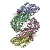
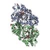
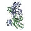
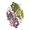
 Vibrio cholerae O1 biovar eltor str. N16961 (bacteria)
 X-RAY DIFFRACTION /
  Authors
 Citation
 Structure visualization
 Downloads & links
 Other downloads
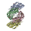


 PDBj
PDBj


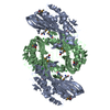
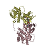
 Assembly
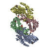


 Mass: 94.971 Da / Num. of mol.: 6 / Source method: obtained synthetically / Formula: PO4
 Mass: 24.305 Da / Num. of mol.: 2 / Source method: obtained synthetically / Formula: Mg
 Mass: 151.126 Da / Num. of mol.: 4 / Source method: obtained synthetically / Formula: C5H5N5O
 Mass: 175.959 Da / Num. of mol.: 2 / Source method: obtained synthetically / Formula: H2O7P2
 Sample preparation
 / Beamline: 19-BM / Wavelength: 0.9793 Å
 Processing