 Movie
Movie Controller

[English] 日本語
 Yorodumi
- PDB-2nxo: Crystal structure of protein SCO4506 from Streptomyces coelicolor... -
+ Open data

- Basic information
| Entry | Database: PDB / ID: 2nxo | ||||||
|---|---|---|---|---|---|---|---|
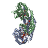
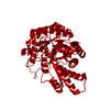
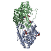
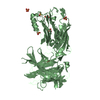
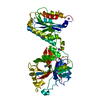
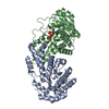
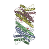
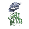
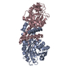
| Title | Crystal structure of protein SCO4506 from Streptomyces coelicolor, Pfam DUF178 | ||||||
 Components Components | Hypothetical protein SCO4506 | ||||||
 Keywords Keywords | STRUCTURAL GENOMICS / UNKNOWN FUNCTION / Pfam / DUf178 / NYSGXRC / 10093f / PSI-2 / Protein Structure Initiative / New York SGX Research Center for Structural Genomics | ||||||
| Function / homology |  Function and homology information Function and homology informationchorismate dehydratase / menaquinone biosynthetic process / hydro-lyase activity Similarity search - Function | ||||||
| Biological species |  Streptomyces coelicolor (bacteria) Streptomyces coelicolor (bacteria) | ||||||
| Method |  X-RAY DIFFRACTION / SYNCHROTRON / SAD / Resolution: 2.04 Å X-RAY DIFFRACTION / SYNCHROTRON / SAD / Resolution: 2.04 Å | ||||||
 Authors Authors | Tyagi, R. / Eswaramoorthy, S. / Burley, S.K. / Swaminathan, S. / New York SGX Research Center for Structural Genomics (NYSGXRC) | ||||||
 Citation Citation | Journal: To be Published Title: The crystal structure of a hypothetical protein SCO4506 (gene ID: Q9L0T8) from Streptomyces coelicolor to 2.04 Angstrom resolution Authors: Tyagi, R. / Eswaramoorthy, S. / Burley, S.K. / Swaminathan, S. | ||||||
| History |
|
- Structure visualization
| Structure viewer | Molecule: MolmilJmol/JSmol |
|---|
- Downloads & links
-Download
| PDBx/mmCIF format | 2nxo.cif.gz | 220.2 KB | Display | PDBx/mmCIF format |
|---|---|---|---|---|
| PDB format | pdb2nxo.ent.gz | 177.7 KB | Display | PDB format |
| PDBx/mmJSON format | 2nxo.json.gz | Tree view | PDBx/mmJSON format | |
| Others |  Other downloads Other downloads |
-Validation report
| Arichive directory | https://data.pdbj.org/pub/pdb/validation_reports/nx/2nxoftp://data.pdbj.org/pub/pdb/validation_reports/nx/2nxo | HTTPS FTP |
|---|
-Related structure data
| Similar structure data | |
|---|---|
| Other databases |

-Links
 PDBj
PDBj- Assembly
| Deposited unit | 
| ||||||||
|---|---|---|---|---|---|---|---|---|---|
| 1 | 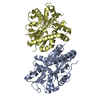
| ||||||||
| 2 | 
| ||||||||
| Unit cell |
|
-Components
| #1: Protein | Mass: 32474.861 Da / Num. of mol.: 4 Source method: isolated from a genetically manipulated source Source: (gene. exp.) Streptomyces coelicolor (bacteria) / Gene: Q9L0T8 / Plasmid: T7 / Production host: #2: Water | ChemComp-HOH / |  Mass: 18.015 Da / Num. of mol.: 550 / Source method: isolated from a natural source / Formula: H2O Mass: 18.015 Da / Num. of mol.: 550 / Source method: isolated from a natural source / Formula: H2O |
|---|
-Experimental details
-Experiment
| Experiment | Method: X-RAY DIFFRACTION / Number of used crystals: 2 |
|---|
- Sample preparation
| Crystal | Density Matthews: 2.36 Å3/Da / Density % sol: 47.81 % |
|---|---|
| Crystal grow | Temperature: 294 K / Method: vapor diffusion, hanging drop / pH: 5.5 Details: 0.2 M MgCl2, 0.1 M Bis-Tris, 25% PEG 3350, pH 5.5, VAPOR DIFFUSION, HANGING DROP, temperature 294K |
-Data collection
| Diffraction |
| ||||||||||||||||||
|---|---|---|---|---|---|---|---|---|---|---|---|---|---|---|---|---|---|---|---|
| Diffraction source |
| ||||||||||||||||||
| Detector |
| ||||||||||||||||||
| Radiation |
| ||||||||||||||||||
| Radiation wavelength |
| ||||||||||||||||||
| Reflection | Resolution: 2.04→50 Å / Num. all: 76032 / Num. obs: 76032 / % possible obs: 98.8 % / Observed criterion σ(I): 0 / Redundancy: 4.6 % / Biso Wilson estimate: 16 Å2 / Rmerge(I) obs: 0.043 / Net I/σ(I): 16.8 | ||||||||||||||||||
| Reflection shell | Resolution: 2.04→2.11 Å / Redundancy: 3.9 % / Rmerge(I) obs: 0.209 / Num. unique all: 6908 / % possible all: 89.7 |

- Processing
| Software |
| |||||||||||||||||||||||||
|---|---|---|---|---|---|---|---|---|---|---|---|---|---|---|---|---|---|---|---|---|---|---|---|---|---|---|
| Refinement | Method to determine structure: SAD / Resolution: 2.04→30.28 Å / Rfactor Rfree error: 0.006 / Data cutoff high absF: 81815.59 / Data cutoff low absF: 0 / Isotropic thermal model: RESTRAINED / Cross valid method: THROUGHOUT / σ(F): 0 / Stereochemistry target values: Engh & Huber Details: The missing residues listed in Remark 465 are due to lack of density. Residues listed having missing atoms in Remark 470 were modeled as ALA due to weak or lack of electron density. ...Details: The missing residues listed in Remark 465 are due to lack of density. Residues listed having missing atoms in Remark 470 were modeled as ALA due to weak or lack of electron density. Continuous residual density in the electron density map which may be due to unidentifiable substrate was modeled with a few waters. This was present in all four monomers.
| |||||||||||||||||||||||||
| Solvent computation | Solvent model: FLAT MODEL / Bsol: 42.1264 Å2 / ksol: 0.328717 e/Å3 | |||||||||||||||||||||||||
| Displacement parameters | Biso mean: 32 Å2
| |||||||||||||||||||||||||
| Refine analyze |
| |||||||||||||||||||||||||
| Refinement step | Cycle: LAST / Resolution: 2.04→30.28 Å
| |||||||||||||||||||||||||
| Refine LS restraints |
| |||||||||||||||||||||||||
| LS refinement shell | Resolution: 2.04→2.17 Å / Rfactor Rfree error: 0.019 / Total num. of bins used: 6
| |||||||||||||||||||||||||
| Xplor file |
|
