 Movie
Movie Controller

[English] 日本語
 Yorodumi
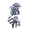
- PDB-4o4x: Crystal structure of the vaccine antigen Transferrin Binding Prot... -
+ Open data

- Basic information
| Entry | Database: PDB / ID: 4o4x | ||||||
|---|---|---|---|---|---|---|---|
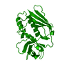
| Title | Crystal structure of the vaccine antigen Transferrin Binding Protein B (TbpB) double mutant Tyr-167-Ala and Trp-176-Ala from Haemophilus parasuis Hp5 | ||||||
 Components Components | TbpB | ||||||
 Keywords Keywords | METAL TRANSPORT / Structure-based vaccine design / transferrin receptor / iron acquisition / host pathogen interaction / surface lipoprotein | ||||||
| Function / homology |  Function and homology information Function and homology information | ||||||
| Biological species |  Haemophilus parasuis (bacteria) Haemophilus parasuis (bacteria) | ||||||
| Method |  X-RAY DIFFRACTION / SYNCHROTRON / MOLECULAR REPLACEMENT / Resolution: 2.9 Å X-RAY DIFFRACTION / SYNCHROTRON / MOLECULAR REPLACEMENT / Resolution: 2.9 Å | ||||||
 Authors Authors | Calmettes, C. / Yu, R.H. / Schryvers, A.B. / Moraes, T.F. | ||||||
 Citation Citation | Journal: Infect.Immun. / Year: 2015 Title: Nonbinding site-directed mutants of transferrin binding protein B exhibit enhanced immunogenicity and protective capabilities. Authors: Frandoloso, R. / Martinez-Martinez, S. / Calmettes, C. / Fegan, J. / Costa, E. / Curran, D. / Yu, R.H. / Gutierrez-Martin, C.B. / Rodriguez-Ferri, E.F. / Moraes, T.F. / Schryvers, A.B. | ||||||
| History |
|
- Structure visualization
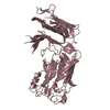
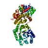
| Structure viewer | Molecule: MolmilJmol/JSmol |
|---|
- Downloads & links
-Download
| PDBx/mmCIF format | 4o4x.cif.gz | 207.6 KB | Display | PDBx/mmCIF format |
|---|---|---|---|---|
| PDB format | pdb4o4x.ent.gz | 165.9 KB | Display | PDB format |
| PDBx/mmJSON format | 4o4x.json.gz | Tree view | PDBx/mmJSON format | |
| Others |  Other downloads Other downloads |
-Validation report
| Arichive directory | https://data.pdbj.org/pub/pdb/validation_reports/o4/4o4xftp://data.pdbj.org/pub/pdb/validation_reports/o4/4o4x | HTTPS FTP |
|---|
-Related structure data
| Related structure data |  4o3wC  4o3xC  4o3yC 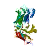 4o3zC  4o49C  4o4uC C: citing same article ( |
|---|---|
| Similar structure data |


-Links
 PDBj
PDBj

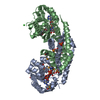
- Assembly
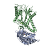
| Deposited unit | 
| ||||||||
|---|---|---|---|---|---|---|---|---|---|
| 1 | 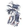
| ||||||||
| 2 | 
| ||||||||
| Unit cell |
|
-Components
| #1: Protein | Mass: 58295.594 Da / Num. of mol.: 2 Source method: isolated from a genetically manipulated source Source: (gene. exp.) Haemophilus parasuis (bacteria) / Plasmid: pET / Production host: #2: Chemical | ChemComp-GOL /   Mass: 92.094 Da / Num. of mol.: 9 / Source method: obtained synthetically / Formula: C3H8O3 Mass: 92.094 Da / Num. of mol.: 9 / Source method: obtained synthetically / Formula: C3H8O3#3: Chemical | ChemComp-SO4 /   Mass: 96.063 Da / Num. of mol.: 10 / Source method: obtained synthetically / Formula: SO4 Mass: 96.063 Da / Num. of mol.: 10 / Source method: obtained synthetically / Formula: SO4#4: Chemical | ChemComp-NA / |   Mass: 22.990 Da / Num. of mol.: 1 / Source method: obtained synthetically / Formula: Na Mass: 22.990 Da / Num. of mol.: 1 / Source method: obtained synthetically / Formula: Na#5: Water | ChemComp-HOH / |  Mass: 18.015 Da / Num. of mol.: 50 / Source method: isolated from a natural source / Formula: H2O Mass: 18.015 Da / Num. of mol.: 50 / Source method: isolated from a natural source / Formula: H2OHas protein modification | Y | |
|---|
-Experimental details
-Experiment
| Experiment | Method: X-RAY DIFFRACTION / Number of used crystals: 1 |
|---|
- Sample preparation
| Crystal | Density Matthews: 2.48 Å3/Da / Density % sol: 50.31 % |
|---|---|
| Crystal grow | Temperature: 293 K / Method: vapor diffusion, hanging drop / pH: 6.5 Details: 0.08 M potassium phosphate monobasic, 18% PEG 8000, 20% glycerol, pH 6.5, VAPOR DIFFUSION, HANGING DROP, temperature 293K |
-Data collection
| Diffraction | Mean temperature: 100 K |
|---|---|
| Diffraction source | Source: SYNCHROTRON / Site: CLSI  / Beamline: 08ID-1 / Wavelength: 0.9795 Å / Beamline: 08ID-1 / Wavelength: 0.9795 Å |
| Detector | Detector: CCD / Date: Jul 7, 2013 |
| Radiation | Protocol: SINGLE WAVELENGTH / Monochromatic (M) / Laue (L): M / Scattering type: x-ray |
| Radiation wavelength | Wavelength: 0.9795 Å / Relative weight: 1 |
| Reflection | Resolution: 2.9→42.182 Å / Num. obs: 26504 / Biso Wilson estimate: 57.75 Å2 |
- Processing
| Software |
| ||||||||||||||||||||||||||||||||||||||||||||||||||||||||||||||||||||||
|---|---|---|---|---|---|---|---|---|---|---|---|---|---|---|---|---|---|---|---|---|---|---|---|---|---|---|---|---|---|---|---|---|---|---|---|---|---|---|---|---|---|---|---|---|---|---|---|---|---|---|---|---|---|---|---|---|---|---|---|---|---|---|---|---|---|---|---|---|---|---|---|
| Refinement | Method to determine structure: MOLECULAR REPLACEMENT / Resolution: 2.9→42.182 Å / Occupancy max: 1 / Occupancy min: 0.4 / SU ML: 0.41 / σ(F): 1.35 / Phase error: 24.09 / Stereochemistry target values: ML
| ||||||||||||||||||||||||||||||||||||||||||||||||||||||||||||||||||||||
| Solvent computation | Shrinkage radii: 0.9 Å / VDW probe radii: 1.11 Å / Solvent model: FLAT BULK SOLVENT MODEL | ||||||||||||||||||||||||||||||||||||||||||||||||||||||||||||||||||||||
| Displacement parameters | Biso mean: 55.5643 Å2 | ||||||||||||||||||||||||||||||||||||||||||||||||||||||||||||||||||||||
| Refinement step | Cycle: LAST / Resolution: 2.9→42.182 Å
| ||||||||||||||||||||||||||||||||||||||||||||||||||||||||||||||||||||||
| Refine LS restraints |
| ||||||||||||||||||||||||||||||||||||||||||||||||||||||||||||||||||||||
| LS refinement shell |
|
