 Movie
Movie Controller

[English] 日本語
 Yorodumi
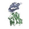
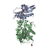
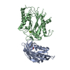
- PDB-3nmv: Crystal structure of pyrabactin-bound abscisic acid receptor PYL2... -
+ Open data

- Basic information
| Entry | Database: PDB / ID: 3nmv | ||||||
|---|---|---|---|---|---|---|---|
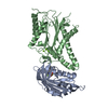
| Title | Crystal structure of pyrabactin-bound abscisic acid receptor PYL2 mutant A93F in complex with type 2C protein phosphatase ABI2 | ||||||
 Components Components |
| ||||||
 Keywords Keywords | PROTEIN BINDING / PYL2 / Pyrabactin / Abscisic acid receptor / helix-grip fold / type 2C protein phosphatase | ||||||
| Function / homology |  Function and homology information Function and homology informationnegative regulation of abscisic acid-activated signaling pathway / regulation of stomatal opening / photoinhibition / protein serine/threonine phosphatase complex / protein phosphatase inhibitor complex / regulation of intracellular signal transduction / abscisic acid binding / response to water deprivation / response to abscisic acid / abscisic acid-activated signaling pathway ...negative regulation of abscisic acid-activated signaling pathway / regulation of stomatal opening / photoinhibition / protein serine/threonine phosphatase complex / protein phosphatase inhibitor complex / regulation of intracellular signal transduction / abscisic acid binding / response to water deprivation / response to abscisic acid / abscisic acid-activated signaling pathway / response to osmotic stress / protein dephosphorylation / protein phosphatase inhibitor activity / protein-serine/threonine phosphatase / protein serine/threonine phosphatase activity / protein kinase activator activity / response to heat / signaling receptor activity / protein homodimerization activity / metal ion binding / identical protein binding / nucleus / plasma membrane / cytoplasm Similarity search - Function | ||||||
| Biological species |  | ||||||
| Method |  X-RAY DIFFRACTION / SYNCHROTRON / MOLECULAR REPLACEMENT / Resolution: 2.1 Å X-RAY DIFFRACTION / SYNCHROTRON / MOLECULAR REPLACEMENT / Resolution: 2.1 Å | ||||||
 Authors Authors | Zhou, X.E. / Melcher, K. / Ng, L.-M. / Soon, F.-F. / Xu, Y. / Suino-Powell, K.M. / Kovach, A. / Li, J. / Yong, E.-L. / Xu, H.E. | ||||||
 Citation Citation | Journal: Nat.Struct.Mol.Biol. / Year: 2010 Title: Identification and mechanism of ABA receptor antagonism. Authors: Melcher, K. / Xu, Y. / Ng, L.M. / Zhou, X.E. / Soon, F.F. / Chinnusamy, V. / Suino-Powell, K.M. / Kovach, A. / Tham, F.S. / Cutler, S.R. / Li, J. / Yong, E.L. / Zhu, J.K. / Xu, H.E. | ||||||
| History |
|
- Structure visualization
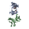
| Structure viewer | Molecule: MolmilJmol/JSmol |
|---|
- Downloads & links
-Download
| PDBx/mmCIF format | 3nmv.cif.gz | 202.3 KB | Display | PDBx/mmCIF format |
|---|---|---|---|---|
| PDB format | pdb3nmv.ent.gz | 159.9 KB | Display | PDB format |
| PDBx/mmJSON format | 3nmv.json.gz | Tree view | PDBx/mmJSON format | |
| Others |  Other downloads Other downloads |
-Validation report
| Arichive directory | https://data.pdbj.org/pub/pdb/validation_reports/nm/3nmvftp://data.pdbj.org/pub/pdb/validation_reports/nm/3nmv | HTTPS FTP |
|---|
-Related structure data
| Related structure data |  3nmhC  3nmnC  3nmpC 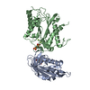 3nmtSC C: citing same article ( S: Starting model for refinement |
|---|---|
| Similar structure data |


-Links
 PDBj
PDBj- Assembly
| Deposited unit | 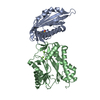
| ||||||||
|---|---|---|---|---|---|---|---|---|---|
| 1 |
| ||||||||
| Unit cell |
|
-Components
| #1: Protein | Mass: 20096.600 Da / Num. of mol.: 1 / Mutation: A93F Source method: isolated from a genetically manipulated source Source: (gene. exp.)  | ||
|---|---|---|---|
| #2: Protein | Mass: 35783.980 Da / Num. of mol.: 1 Source method: isolated from a genetically manipulated source Source: (gene. exp.) References: UniProt: O04719, protein-serine/threonine phosphatase | ||
| #3: Chemical | ChemComp-PYV / 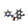  Mass: 377.256 Da / Num. of mol.: 1 / Source method: obtained synthetically / Formula: C16H13BrN2O2S / Comment: hormone*YM Mass: 377.256 Da / Num. of mol.: 1 / Source method: obtained synthetically / Formula: C16H13BrN2O2S / Comment: hormone*YM | ||
| #4: Chemical |   Mass: 24.305 Da / Num. of mol.: 3 / Source method: obtained synthetically / Formula: Mg Mass: 24.305 Da / Num. of mol.: 3 / Source method: obtained synthetically / Formula: Mg#5: Water | ChemComp-HOH / |  Mass: 18.015 Da / Num. of mol.: 171 / Source method: isolated from a natural source / Formula: H2O Mass: 18.015 Da / Num. of mol.: 171 / Source method: isolated from a natural source / Formula: H2O |
-Experimental details
-Experiment
| Experiment | Method: X-RAY DIFFRACTION / Number of used crystals: 1 |
|---|
- Sample preparation
| Crystal | Density Matthews: 3.65 Å3/Da / Density % sol: 66.29 % |
|---|---|
| Crystal grow | Temperature: 293 K / Method: vapor diffusion, hanging drop / pH: 7.5 Details: 0.1M HEPES, 10% PEG 8000, 10% sucrose, pH 7.5, VAPOR DIFFUSION, HANGING DROP, temperature 293K |
-Data collection
| Diffraction | Mean temperature: 100 K |
|---|---|
| Diffraction source | Source: SYNCHROTRON / Site: APS  / Beamline: 21-ID-D / Wavelength: 1 Å / Beamline: 21-ID-D / Wavelength: 1 Å |
| Detector | Type: MARMOSAIC 300 mm CCD / Detector: CCD |
| Radiation | Protocol: SINGLE WAVELENGTH / Monochromatic (M) / Laue (L): M / Scattering type: x-ray |
| Radiation wavelength | Wavelength: 1 Å / Relative weight: 1 |
| Reflection | Resolution: 2.1→30 Å / Num. obs: 44745 / % possible obs: 98.2 % / Observed criterion σ(F): 2 / Observed criterion σ(I): 2 / Redundancy: 14.7 % / Rmerge(I) obs: 0.1 / Net I/σ(I): 32.4 |
- Processing
| Software |
| ||||||||||||||||||||||||||||||||||||||||||||||||||||||||||||||||||||||||||||||||||||||||||||||||||||||||||||||||||||||||||||||||||||||||||||||||||||||||||||||||||||||||||
|---|---|---|---|---|---|---|---|---|---|---|---|---|---|---|---|---|---|---|---|---|---|---|---|---|---|---|---|---|---|---|---|---|---|---|---|---|---|---|---|---|---|---|---|---|---|---|---|---|---|---|---|---|---|---|---|---|---|---|---|---|---|---|---|---|---|---|---|---|---|---|---|---|---|---|---|---|---|---|---|---|---|---|---|---|---|---|---|---|---|---|---|---|---|---|---|---|---|---|---|---|---|---|---|---|---|---|---|---|---|---|---|---|---|---|---|---|---|---|---|---|---|---|---|---|---|---|---|---|---|---|---|---|---|---|---|---|---|---|---|---|---|---|---|---|---|---|---|---|---|---|---|---|---|---|---|---|---|---|---|---|---|---|---|---|---|---|---|---|---|---|---|
| Refinement | Method to determine structure: MOLECULAR REPLACEMENT Starting model: PDB entry 3NMT Resolution: 2.1→29.6 Å / Cor.coef. Fo:Fc: 0.955 / Cor.coef. Fo:Fc free: 0.945 / SU B: 8.592 / SU ML: 0.102 / Cross valid method: THROUGHOUT / ESU R Free: 0.14 / Stereochemistry target values: MAXIMUM LIKELIHOOD / Details: HYDROGENS HAVE BEEN ADDED IN THE RIDING POSITIONS
| ||||||||||||||||||||||||||||||||||||||||||||||||||||||||||||||||||||||||||||||||||||||||||||||||||||||||||||||||||||||||||||||||||||||||||||||||||||||||||||||||||||||||||
| Solvent computation | Ion probe radii: 0.8 Å / Shrinkage radii: 0.8 Å / VDW probe radii: 1.4 Å / Solvent model: BABINET MODEL WITH MASK | ||||||||||||||||||||||||||||||||||||||||||||||||||||||||||||||||||||||||||||||||||||||||||||||||||||||||||||||||||||||||||||||||||||||||||||||||||||||||||||||||||||||||||
| Displacement parameters | Biso mean: 38.284 Å2
| ||||||||||||||||||||||||||||||||||||||||||||||||||||||||||||||||||||||||||||||||||||||||||||||||||||||||||||||||||||||||||||||||||||||||||||||||||||||||||||||||||||||||||
| Refinement step | Cycle: LAST / Resolution: 2.1→29.6 Å
| ||||||||||||||||||||||||||||||||||||||||||||||||||||||||||||||||||||||||||||||||||||||||||||||||||||||||||||||||||||||||||||||||||||||||||||||||||||||||||||||||||||||||||
| Refine LS restraints |
| ||||||||||||||||||||||||||||||||||||||||||||||||||||||||||||||||||||||||||||||||||||||||||||||||||||||||||||||||||||||||||||||||||||||||||||||||||||||||||||||||||||||||||
| LS refinement shell | Resolution: 2.1→2.158 Å / Total num. of bins used: 20
|
