 Movie
Movie Controller

[English] 日本語
 Yorodumi
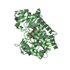
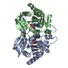
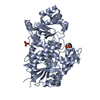
- PDB-2nrf: Crystal Structure of GlpG, a Rhomboid family intramembrane protease -
+ Open data

- Basic information
| Entry | Database: PDB / ID: 2nrf | ||||||
|---|---|---|---|---|---|---|---|
| Title | Crystal Structure of GlpG, a Rhomboid family intramembrane protease | ||||||
 Components Components | Protein GlpG | ||||||
 Keywords Keywords | MEMBRANE PROTEIN / integral membrane protein | ||||||
| Function / homology |  Function and homology information Function and homology informationrhomboid protease / endopeptidase activity / serine-type endopeptidase activity / proteolysis / identical protein binding / plasma membrane Similarity search - Function | ||||||
| Biological species |  | ||||||
| Method |  X-RAY DIFFRACTION / SYNCHROTRON / MOLECULAR REPLACEMENT / Resolution: 2.6 Å X-RAY DIFFRACTION / SYNCHROTRON / MOLECULAR REPLACEMENT / Resolution: 2.6 Å | ||||||
 Authors Authors | Wu, Z. / Yan, N. / Feng, L. / Yan, H. / Gu, L. / Shi, Y. | ||||||
 Citation Citation | Journal: Nat.Struct.Mol.Biol. / Year: 2006 Title: Structural analysis of a rhomboid family intramembrane protease reveals a gating mechanism for substrate entry. Authors: Wu, Z. / Yan, N. / Feng, L. / Oberstein, A. / Yan, H. / Baker, R.P. / Gu, L. / Jeffrey, P.D. / Urban, S. / Shi, Y. | ||||||
| History |
|
- Structure visualization
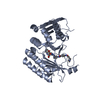
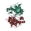
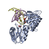
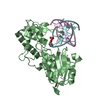
| Structure viewer | Molecule: MolmilJmol/JSmol |
|---|
- Downloads & links
-Download
| PDBx/mmCIF format | 2nrf.cif.gz | 81.7 KB | Display | PDBx/mmCIF format |
|---|---|---|---|---|
| PDB format | pdb2nrf.ent.gz | 62 KB | Display | PDB format |
| PDBx/mmJSON format | 2nrf.json.gz | Tree view | PDBx/mmJSON format | |
| Others |  Other downloads Other downloads |
-Validation report
| Arichive directory | https://data.pdbj.org/pub/pdb/validation_reports/nr/2nrfftp://data.pdbj.org/pub/pdb/validation_reports/nr/2nrf | HTTPS FTP |
|---|
-Related structure data
| Related structure data | 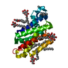 2ic8S S: Starting model for refinement |
|---|---|
| Similar structure data |


-Links
 PDBj
PDBj
- Assembly
| Deposited unit | 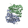
| ||||||||
|---|---|---|---|---|---|---|---|---|---|
| 1 |
| ||||||||
| Unit cell |
| ||||||||
| Details | Each asymmetric unit contains two molecules. They exhibit different conformations. |
-Components
| #1: Protein | Mass: 20528.312 Da / Num. of mol.: 2 Source method: isolated from a genetically manipulated source Source: (gene. exp.) #2: Water | ChemComp-HOH / |  Mass: 18.015 Da / Num. of mol.: 26 / Source method: isolated from a natural source / Formula: H2O Mass: 18.015 Da / Num. of mol.: 26 / Source method: isolated from a natural source / Formula: H2O |
|---|
-Experimental details
-Experiment
| Experiment | Method: X-RAY DIFFRACTION / Number of used crystals: 1 |
|---|
- Sample preparation
| Crystal | Density Matthews: 2.93 Å3/Da / Density % sol: 58.07 % |
|---|---|
| Crystal grow | Temperature: 295 K / Method: vapor diffusion, hanging drop / pH: 7.4 Details: PEG 3000 7%, Li2S04 100 mM, pH 7.4, VAPOR DIFFUSION, HANGING DROP, temperature 295K |
-Data collection
| Diffraction | Mean temperature: 100 K |
|---|---|
| Diffraction source | Source: SYNCHROTRON / Site: NSLS  / Beamline: X29A / Wavelength: 0.9793 Å / Beamline: X29A / Wavelength: 0.9793 Å |
| Detector | Type: ADSC QUANTUM 315 / Detector: CCD / Date: Sep 13, 2006 |
| Radiation | Monochromator: a vertically focusing mirror and a horizontally focusing monochromator Protocol: SINGLE WAVELENGTH / Monochromatic (M) / Laue (L): M / Scattering type: x-ray |
| Radiation wavelength | Wavelength: 0.9793 Å / Relative weight: 1 |
| Reflection twin | Type: hemihedral / Operator: h,-h-k,-l / Fraction: 0.4 |
| Reflection | Resolution: 2.6→100 Å / Num. all: 14363 / Num. obs: 14184 / % possible obs: 98.5 % / Observed criterion σ(F): 0 / Observed criterion σ(I): 1 / Redundancy: 9 % / Rmerge(I) obs: 0.071 / Χ2: 1.033 / Net I/σ(I): 15.1 |
| Reflection shell | Resolution: 2.6→2.69 Å / Redundancy: 6.3 % / Rmerge(I) obs: 0.508 / Num. unique all: 1373 / Χ2: 0.446 / % possible all: 93.9 |
- Processing
| Software |
| ||||||||||||||||||||||||||||
|---|---|---|---|---|---|---|---|---|---|---|---|---|---|---|---|---|---|---|---|---|---|---|---|---|---|---|---|---|---|
| Refinement | Method to determine structure: MOLECULAR REPLACEMENT Starting model: PDB ENTRY 2IC8 Resolution: 2.6→100 Å / Cross valid method: THROUGHOUT / σ(F): 0 / Stereochemistry target values: Engh & Huber
| ||||||||||||||||||||||||||||
| Solvent computation | Bsol: 90.08 Å2 | ||||||||||||||||||||||||||||
| Displacement parameters | Biso mean: 102.891 Å2
| ||||||||||||||||||||||||||||
| Refinement step | Cycle: LAST / Resolution: 2.6→100 Å
| ||||||||||||||||||||||||||||
| Refine LS restraints |
| ||||||||||||||||||||||||||||
| LS refinement shell | Resolution: 2.6→50 Å | ||||||||||||||||||||||||||||
| Xplor file |
|
