 Movie
Movie Controller

[English] 日本語
 Yorodumi
- PDB-2nlr: STREPTOMYCES LIVIDANS ENDOGLUCANASE (EC: 3.2.1.4) COMPLEX WITH MO... -
+ Open data

- Basic information
| Entry | Database: PDB / ID: 2nlr | |||||||||
|---|---|---|---|---|---|---|---|---|---|---|
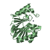
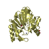
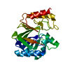
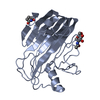
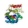
| Title | STREPTOMYCES LIVIDANS ENDOGLUCANASE (EC: 3.2.1.4) COMPLEX WITH MODIFIED GLUCOSE TRIMER | |||||||||
 Components Components | PROTEIN (ENDOGLUCANASE (E.C.3.2.1.4)) | |||||||||
 Keywords Keywords | HYDROLASE / HYDROLASE (ENDOGLUCANASE) / GLYCOSYL HYDROLASE / FAMILY 12 / ENDOGLUCANASE / CELB2 / GLYCOSYL-ENZYME INTERMEDIATE | |||||||||
| Function / homology |  Function and homology information Function and homology informationcellulase activity / polysaccharide binding / polysaccharide catabolic process Similarity search - Function | |||||||||
| Biological species |  Streptomyces lividans (bacteria) Streptomyces lividans (bacteria) | |||||||||
| Method |  X-RAY DIFFRACTION / SYNCHROTRON / MOLECULAR REPLACEMENT / Resolution: 1.2 Å X-RAY DIFFRACTION / SYNCHROTRON / MOLECULAR REPLACEMENT / Resolution: 1.2 Å | |||||||||
 Authors Authors | Sulzenbacher, G. / Dupont, C. / Davies, G.J. | |||||||||
 Citation Citation | Journal: Biochemistry / Year: 1999 Title: The crystal structure of a 2-fluorocellotriosyl complex of the Streptomyces lividans endoglucanase CelB2 at 1.2 A resolution. Authors: Sulzenbacher, G. / Mackenzie, L.F. / Wilson, K.S. / Withers, S.G. / Dupont, C. / Davies, G.J. #1: Journal: Biochemistry / Year: 1997Title: The Streptomyces lividans Family 12 Endoglucanase: Construction of the Catalytic Core, Expression and X-Ray Structure at 1.75 A Resolution Authors: Sulzenbacher, G. / Sharek, F. / Morosoli, R. / Dupont, C. / Davies, G.J. #2: Journal: Appl.Environ.Microbiol. / Year: 1994Title: Purification and Characterisation of the Celb Endoglucanase from Streptomyces lividans 66 and DNA Sequence of the Encoding Gene Authors: Wittmann, S. / Shareck, F. / Kluepfel, D. / Morosoli, R. | |||||||||
| History |
|
- Structure visualization
| Structure viewer | Molecule: MolmilJmol/JSmol |
|---|
- Downloads & links
-Download
| PDBx/mmCIF format | 2nlr.cif.gz | 179 KB | Display | PDBx/mmCIF format |
|---|---|---|---|---|
| PDB format | pdb2nlr.ent.gz | 140.1 KB | Display | PDB format |
| PDBx/mmJSON format | 2nlr.json.gz | Tree view | PDBx/mmJSON format | |
| Others |  Other downloads Other downloads |
-Validation report
| Arichive directory | https://data.pdbj.org/pub/pdb/validation_reports/nl/2nlrftp://data.pdbj.org/pub/pdb/validation_reports/nl/2nlr | HTTPS FTP |
|---|
-Related structure data
| Related structure data |  1nlrS S: Starting model for refinement |
|---|---|
| Similar structure data |


-Links
 PDBj
PDBj

- Assembly
| Deposited unit | 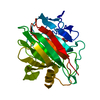
| ||||||||
|---|---|---|---|---|---|---|---|---|---|
| 1 |
| ||||||||
| Unit cell |
| ||||||||
| Components on special symmetry positions |
|
-Components
| #1: Protein | Mass: 24578.713 Da / Num. of mol.: 1 / Fragment: CATALYTIC DOMAIN Source method: isolated from a genetically manipulated source Source: (gene. exp.) Streptomyces lividans (bacteria) / Strain: 66 / Gene: CELB / Plasmid: PIAF9, PIAF18 / Production host: Streptomyces lividans (bacteria) / Strain (production host): 66References: GenBank: 2462718, UniProt: Q54331*PLUS, cellulase |
|---|---|
| #2: Polysaccharide | beta-D-glucopyranose-(1-4)-beta-D-glucopyranose-(1-4)-2-deoxy-2-fluoro-beta-D-glucopyranose Source method: isolated from a genetically manipulated source |
| #3: Water | ChemComp-HOH /  Mass: 18.015 Da / Num. of mol.: 270 / Source method: isolated from a natural source / Formula: H2O Mass: 18.015 Da / Num. of mol.: 270 / Source method: isolated from a natural source / Formula: H2O |
| Compound details | ENDOGLUCANASE CELB BELONGS TO GLYCOSYL HYDROLASASE FAMILY 12. THE ENZYME PERFORMS CATALYSIS WITH ...ENDOGLUCAN |
| Has protein modification | Y |
| Sequence details | THE CONSTRUCT CELB2 WAS AMPLIFIED UP TO RESIDUE 274 (U04629 WHICH IS THE END OF THE JUNCTION ...THE CONSTRUCT CELB2 WAS AMPLIFIED UP TO RESIDUE 274 (U04629 WHICH IS THE END OF THE JUNCTION BETWEEN THE CATALYTIC DOMAIN AND THE CELLULOSE BINDING DOMAIN OF CELB. THE DEPOSITED COORDINATE |
-Experimental details
-Experiment
| Experiment | Method: X-RAY DIFFRACTION / Number of used crystals: 1 |
|---|
- Sample preparation
| Crystal | Density Matthews: 2.34 Å3/Da / Density % sol: 47 % | ||||||||||||||||||||
|---|---|---|---|---|---|---|---|---|---|---|---|---|---|---|---|---|---|---|---|---|---|
| Crystal grow | pH: 4.5 / Details: pH 4.5 | ||||||||||||||||||||
| Crystal grow | *PLUS Method: vapor diffusion, hanging drop | ||||||||||||||||||||
| Components of the solutions | *PLUS
|
-Data collection
| Diffraction | Mean temperature: 120 K |
|---|---|
| Diffraction source | Source: SYNCHROTRON / Site: EMBL/DESY, HAMBURG  / Beamline: X11 / Beamline: X11 |
| Detector | Type: MARRESEARCH / Detector: IMAGE PLATE / Date: Oct 1, 1997 / Details: FOCUSING MIRRORS |
| Radiation | Monochromator: SI / Protocol: SINGLE WAVELENGTH / Monochromatic (M) / Laue (L): M / Scattering type: x-ray |
| Radiation wavelength | Relative weight: 1 |
| Reflection | Resolution: 1.2→25 Å / Num. obs: 69645 / % possible obs: 99.1 % / Redundancy: 5.96 % / Rmerge(I) obs: 0.054 / Net I/σ(I): 14.93 |
| Reflection shell | Resolution: 1.2→1.22 Å / Redundancy: 4.49 % / Rmerge(I) obs: 0.408 / Mean I/σ(I) obs: 3.74 / % possible all: 97.2 |
| Reflection shell | *PLUS % possible obs: 97.2 % |
- Processing
| Software |
| |||||||||||||||||||||||||||||||||
|---|---|---|---|---|---|---|---|---|---|---|---|---|---|---|---|---|---|---|---|---|---|---|---|---|---|---|---|---|---|---|---|---|---|---|
| Refinement | Method to determine structure: MOLECULAR REPLACEMENT Starting model: PDB ENTRY 1NLR Resolution: 1.2→25 Å / Num. parameters: 20212 / Num. restraintsaints: 28536 / Cross valid method: FREE R / σ(F): 0 StereochEM target val spec case: TARGET VALUES FOR 2-DEOXY-2-FLUORO-CELLOTRIO WERE TAKEN FROM THE CRYSTAL STRUCTURE OF METHYL-BETA-CELL TRIOSIDE; S.RAYMOND, B.HENRISSAT,D.T.QUI,A.KVICK, H.CHANZY, ...StereochEM target val spec case: TARGET VALUES FOR 2-DEOXY-2-FLUORO-CELLOTRIO WERE TAKEN FROM THE CRYSTAL STRUCTURE OF METHYL-BETA-CELL TRIOSIDE; S.RAYMOND, B.HENRISSAT,D.T.QUI,A.KVICK, H.CHANZY, CAROBOHYDRATE RESEARCH 277,209-229. Stereochemistry target values: ENGH AND HUBER Details: ANISOTROPIC REFINEMENT REDUCED FREE R (NO CUTOFF) BY 3.95. RESIDUES ASP 104, GLU 120 AND MET 122 EXHIBIT DISORDER WHICH IS COUPLED TO THE TWO CONFORMATIONS OF 2-DEOXY-2-FLUORO-GLUCOSE. THE ...Details: ANISOTROPIC REFINEMENT REDUCED FREE R (NO CUTOFF) BY 3.95. RESIDUES ASP 104, GLU 120 AND MET 122 EXHIBIT DISORDER WHICH IS COUPLED TO THE TWO CONFORMATIONS OF 2-DEOXY-2-FLUORO-GLUCOSE. THE NUCLEOPHILE GLU 120 IS PRESENT IN TRIPLE CONFORMATION. CONFORMER B AND C ARE COVALENTLY LINKED TO THE FLUOROCELLOTRIOSIDE. HYDROGEN ATOMS HAVE NOT BEEN LOCATED ONLY ON ALL DISORDERED RESIDUES. ASP 95 AND GLY 96 SIT NEAR THE CRYSTALLOGRAPHIC TWOFOLD AXIS AND ARE PRESENT IN TWO ALTERNATIVE AND COMPLEMENTARY CONFORMATIONS. THR 222 IS THE LAST VISIBLE RESIDUE IN THE ELECTRON DENSITY MAP. PRO 76 IS PRESENT IN THE CIS CONFORMATION.
| |||||||||||||||||||||||||||||||||
| Solvent computation | Solvent model: SHELX SWAT | |||||||||||||||||||||||||||||||||
| Refine analyze | Num. disordered residues: 34 / Occupancy sum hydrogen: 1509 / Occupancy sum non hydrogen: 1936.6 | |||||||||||||||||||||||||||||||||
| Refinement step | Cycle: LAST / Resolution: 1.2→25 Å
| |||||||||||||||||||||||||||||||||
| Refine LS restraints |
| |||||||||||||||||||||||||||||||||
| Software | *PLUS Name: SHELXL-96 / Classification: refinement | |||||||||||||||||||||||||||||||||
| Refine LS restraints | *PLUS
|
