 Movie
Movie Controller

[English] 日本語
 Yorodumi
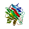
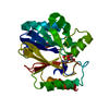
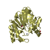
- PDB-5gm4: Crystal structure of FI-CMCase from Aspergillus aculeatus F-50 in... -
+ Open data

- Basic information
| Entry | Database: PDB / ID: 5gm4 | |||||||||
|---|---|---|---|---|---|---|---|---|---|---|
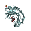
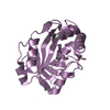
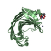
| Title | Crystal structure of FI-CMCase from Aspergillus aculeatus F-50 in complex with cellotetrose | |||||||||
 Components Components | Endoglucanase-1 | |||||||||
 Keywords Keywords | HYDROLASE/INHIBITOR / substrate binding / HYDROLASE-INHIBITOR complex | |||||||||
| Function / homology |  Function and homology information Function and homology informationcellulase / cellulase activity / cellulose catabolic process / extracellular region Similarity search - Function | |||||||||
| Biological species |  | |||||||||
| Method |  X-RAY DIFFRACTION / SYNCHROTRON / MOLECULAR REPLACEMENT / Resolution: 1.92 Å X-RAY DIFFRACTION / SYNCHROTRON / MOLECULAR REPLACEMENT / Resolution: 1.92 Å | |||||||||
 Authors Authors | Huang, J.W. / Liu, W.D. / Zheng, Y.Y. / Chen, C.C. / Guo, R.T. | |||||||||
 Citation Citation | Journal: Biochem. Biophys. Res. Commun. / Year: 2016 Title: Crystal structure and genetic modifications of FI-CMCase from Aspergillus aculeatus F-50 Authors: Huang, J.W. / Liu, W. / Lai, H.L. / Cheng, Y.S. / Zheng, Y. / Li, Q. / Sun, H. / Kuo, C.J. / Guo, R.T. / Chen, C.C. | |||||||||
| History |
|
- Structure visualization
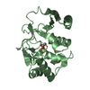
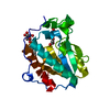
| Structure viewer | Molecule: MolmilJmol/JSmol |
|---|
- Downloads & links
-Download
| PDBx/mmCIF format | 5gm4.cif.gz | 329.9 KB | Display | PDBx/mmCIF format |
|---|---|---|---|---|
| PDB format | pdb5gm4.ent.gz | 268.5 KB | Display | PDB format |
| PDBx/mmJSON format | 5gm4.json.gz | Tree view | PDBx/mmJSON format | |
| Others |  Other downloads Other downloads |
-Validation report
| Arichive directory | https://data.pdbj.org/pub/pdb/validation_reports/gm/5gm4ftp://data.pdbj.org/pub/pdb/validation_reports/gm/5gm4 | HTTPS FTP |
|---|
-Related structure data
| Related structure data | 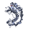 5gm3C 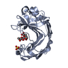 5gm5C  1ks4S C: citing same article ( S: Starting model for refinement |
|---|---|
| Similar structure data |

-Links
 PDBj
PDBj
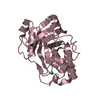
- Assembly
| Deposited unit | 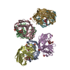
| ||||||||
|---|---|---|---|---|---|---|---|---|---|
| 1 | 
| ||||||||
| 2 | 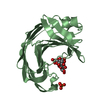
| ||||||||
| 3 | 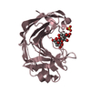
| ||||||||
| 4 | 
| ||||||||
| 5 | 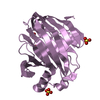
| ||||||||
| 6 | 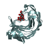
| ||||||||
| 7 | 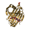
| ||||||||
| Unit cell |
|
-Components
| #1: Protein | Mass: 23720.842 Da / Num. of mol.: 7 / Fragment: UNP residues 19-237 / Mutation: E202A Source method: isolated from a genetically manipulated source Source: (gene. exp.) Pichia pastoris (fungus) / Strain (production host): X33 / References: UniProt: P22669, cellulase#2: Polysaccharide | beta-D-glucopyranose-(1-4)-beta-D-glucopyranose-(1-4)-beta-D-glucopyranose-(1-4)-beta-D-glucopyranose / beta-cellotetraose 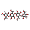  Source method: isolated from a genetically manipulated source Details: oligosaccharide / References: beta-cellotetraose #3: Chemical | ChemComp-SO4 /   Mass: 96.063 Da / Num. of mol.: 8 / Source method: obtained synthetically / Formula: SO4 Mass: 96.063 Da / Num. of mol.: 8 / Source method: obtained synthetically / Formula: SO4#4: Water | ChemComp-HOH / |  Mass: 18.015 Da / Num. of mol.: 1187 / Source method: isolated from a natural source / Formula: H2O Mass: 18.015 Da / Num. of mol.: 1187 / Source method: isolated from a natural source / Formula: H2OHas protein modification | Y | |
|---|
-Experimental details
-Experiment
| Experiment | Method: X-RAY DIFFRACTION / Number of used crystals: 1 |
|---|
- Sample preparation
| Crystal | Density Matthews: 2.39 Å3/Da / Density % sol: 48.56 % / Mosaicity: 0.573 ° |
|---|---|
| Crystal grow | Temperature: 298 K / Method: vapor diffusion, sitting drop / pH: 7.5 / Details: lithium sulfate, HEPES, zinc acetate, |
-Data collection
| Diffraction | Mean temperature: 100 K | ||||||||||||||||||||||||||||||||||||||||||||||||||||||||||||||||||
|---|---|---|---|---|---|---|---|---|---|---|---|---|---|---|---|---|---|---|---|---|---|---|---|---|---|---|---|---|---|---|---|---|---|---|---|---|---|---|---|---|---|---|---|---|---|---|---|---|---|---|---|---|---|---|---|---|---|---|---|---|---|---|---|---|---|---|---|
| Diffraction source | Source: SYNCHROTRON / Site: NSRRC  / Beamline: BL13C1 / Wavelength: 1 Å / Beamline: BL13C1 / Wavelength: 1 Å | ||||||||||||||||||||||||||||||||||||||||||||||||||||||||||||||||||
| Detector | Type: ADSC QUANTUM 315r / Detector: CCD / Date: Mar 18, 2015 | ||||||||||||||||||||||||||||||||||||||||||||||||||||||||||||||||||
| Radiation | Monochromator: GRAPHITE / Protocol: SINGLE WAVELENGTH / Monochromatic (M) / Laue (L): M / Scattering type: x-ray | ||||||||||||||||||||||||||||||||||||||||||||||||||||||||||||||||||
| Radiation wavelength | Wavelength: 1 Å / Relative weight: 1 | ||||||||||||||||||||||||||||||||||||||||||||||||||||||||||||||||||
| Reflection | Resolution: 1.92→25 Å / Num. obs: 118911 / % possible obs: 99.9 % / Redundancy: 4 % / Rmerge(I) obs: 0.065 / Rpim(I) all: 0.037 / Rrim(I) all: 0.075 / Χ2: 1.348 / Net I/av σ(I): 28.671 / Net I/σ(I): 11.6 / Num. measured all: 480843 | ||||||||||||||||||||||||||||||||||||||||||||||||||||||||||||||||||
| Reflection shell |
|
- Processing
| Software |
| |||||||||||||||||||||||||||||||||||||||||||||||||||||||||||||||||||||||||||
|---|---|---|---|---|---|---|---|---|---|---|---|---|---|---|---|---|---|---|---|---|---|---|---|---|---|---|---|---|---|---|---|---|---|---|---|---|---|---|---|---|---|---|---|---|---|---|---|---|---|---|---|---|---|---|---|---|---|---|---|---|---|---|---|---|---|---|---|---|---|---|---|---|---|---|---|---|
| Refinement | Method to determine structure: MOLECULAR REPLACEMENT Starting model: 1KS4 Resolution: 1.92→25 Å / Cor.coef. Fo:Fc: 0.971 / Cor.coef. Fo:Fc free: 0.951 / SU B: 3.435 / SU ML: 0.098 / Cross valid method: THROUGHOUT / σ(F): 0 / ESU R: 0.143 / ESU R Free: 0.139 Details: HYDROGENS HAVE BEEN ADDED IN THE RIDING POSITIONS U VALUES : REFINED INDIVIDUALLY
| |||||||||||||||||||||||||||||||||||||||||||||||||||||||||||||||||||||||||||
| Solvent computation | Ion probe radii: 0.8 Å / Shrinkage radii: 0.8 Å / VDW probe radii: 1.2 Å | |||||||||||||||||||||||||||||||||||||||||||||||||||||||||||||||||||||||||||
| Displacement parameters | Biso max: 111.27 Å2 / Biso mean: 29.236 Å2 / Biso min: 14.97 Å2
| |||||||||||||||||||||||||||||||||||||||||||||||||||||||||||||||||||||||||||
| Refinement step | Cycle: final / Resolution: 1.92→25 Å
| |||||||||||||||||||||||||||||||||||||||||||||||||||||||||||||||||||||||||||
| Refine LS restraints |
| |||||||||||||||||||||||||||||||||||||||||||||||||||||||||||||||||||||||||||
| LS refinement shell | Resolution: 1.922→1.972 Å / Total num. of bins used: 20
|
